This is an automatically translated article.
Herniated disc is a disc that is moved out of position inside the vertebrae, when impacted or traumatized or the disc is degenerated, torn, and clinically often manifests as neuropathic pain .
1. Symptoms of herniated disc
When muscle groups are stimulated, causing severe pain when moving such as: bending, coughing, sneezing or exerting. When sitting for a long time makes the pain worse, either standing or lying on your stomach. Herniated disc in the low back: causes back pain, with or without pain of sciatic nerve symptoms. Herniated disc in the neck: stiffness and pain in the neck. Pain radiating to the shoulders and arms (usually one side), accompanied by a tingling sensation and heaviness or weakness in the legs or arms. Symptoms of numbness of hands and feet: the mucus of the disc coming out will compress the nerve roots causing pain, numbness in the lumbar region, the neck area then gradually develops down the buttocks, thighs, groin and heels. . The patient has sensory disturbances, feeling like ants crawling in the body. Polio, muscle weakness: when it is in the severe stage, it takes a while to be detected. At this stage, the patient can hardly walk, long-term leading to atrophy of the legs, muscle atrophy, paralysis of the limbs, must lie down or sit in a wheelchair.
2. Cause
Some of the main causes of herniated discs that a person may experience are as follows:
Due to work, movement, overwork or wrong posture, leading to disc and spine damage Due to age : is the cause that most patients encounter. When the aging process takes place, the discs and spine become dehydrated, sclerosis degeneration and very easily damaged Due to trauma in the back region Congenital diseases such as or acquired in the spine area such as kyphosis , spondylolisthesis ... Genetic factors In addition, there are several risk factors for herniated disc disease such as:
Body weight: the greater the body weight, the greater the burden on the discs. The higher the spinal cushion, especially in the lumbar region. Occupation: subjects who do manual labor, carry heavy loads, and have poor posture are at high risk of disc herniation.
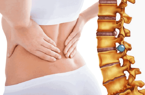
Thoát vị đĩa đệm là đĩa đệm bị di chuyển ra khỏi vị trí bên trong đốt sống
3. Diagnostic Imaging
3.1. Routine X-ray
For X-rays of the spine taken in two upright and inclined positions, Barr's triad can be seen:
+ Scoliosis on straight film.
+ Reduce intervertebral height.
+ Reduce spinal curvature in tilt film.
Because the disc is a non-contrast organ, the image cannot be seen directly, it can only be assessed indirectly through the change of the intervertebral space, adjacent vertebrae and spinal curve.
3.2. Nerve root capsule
Using contrast agent injected into the subarachnoid space of the spinal cord, take two films straight - tilted and 3⁄4 right, left.
On straight film shows the image of nerve root amputation, concave compression of the contrast column, (may have hourglass shape), interruption of the column or complete severing of the contrast column.
This is a method for indirect imaging of disc herniation by imaging spinal stenosis, synaptic foramen, but cannot give direct image of disc, so it cannot distinguish compression caused by other causes. Currently, with the advent of magnetic resonance imaging, nerve root sheath imaging is rarely applied.
3.3. Computed tomography
Valuable in cases of disc herniation with bone degeneration such as calcification of the posterior longitudinal ligament, thickening of the yellow ligament and the beak. However, computed tomography is limited in evaluating disc structure and degree of herniation.
2.4. Magnetic resonance imaging (MRI)
On the magnetic resonance image of the organization, there are many countries with reduced signal on T1 image and increased signal on T2 image. The normal intervertebral disc is well demarcated, hypointensity on T1 and hyperintensity on T2 due to excess fluid. The discs degenerate due to the absence of water, so the signal on T2 is not increased compared to other discs. The herniated disc mass is the part that is co-signal with the disc and protrudes posteriorly from the posterior border of the vertebral body, which is clearly seen on T1- and T2-weighted images. Posterior hernias are the most common, based on longitudinal or transverse images to evaluate the types of hernias. Accurately determine the position of the herniated disc relative to the canal and the degree of spinal cord and nerve root compression.
Magnetic resonance imaging is considered the "gold standard" test in the diagnosis of herniated disc, allowing to exclude lesions inside the spinal cord. Magnetic resonance imaging for canals, disc images with high resolution, observed in many different directions, is a safe and non-toxic method for patients.
4. Treatment
Method of treating herniated disc through the skin by DSA imaging technique:
4.1. Anesthesia method: Local anesthetic with Lidocaine 2% (2-10ml).
4.2. Technique
Put the patient on the table to increase the light. Place an intravenous line. Locate the disc to be treated under the light curtain. Disinfect the injured area. The doctor washes his hands, puts on a shirt, wears gloves, and spreads a sterile canvas with holes on the site to be biopsied. Local anesthetic layer by layer Insert the needle through the skin into the disc to be treated, control the puncture under the light curtain. When the needle is inserted into the right center of the disc nucleus, depending on the treatment purpose, it is possible to inject chemicals, or burn the disc mucus with high-frequency waves. Tape the puncture site.
5. Prevention
Measures to prevent disc herniation can be done as follows:
Exercise with moderate sports, increase the flexibility of the muscles next to the spine. This can help stabilize the spine, reducing the risk of disc damage. Do not carry, exercise too much or have poor posture Maintain a weight appropriate for height, avoid maintaining too much pressure on the spine . When detecting unusual pain in the spine, the patient should immediately go to a medical facility, see a specialist to be accurately diagnosed with the cause and receive timely treatment.
Please dial HOTLINE for more information or register for an appointment HERE. Download MyVinmec app to make appointments faster and to manage your bookings easily.


