This is an automatically translated article.
Article by Doctor Mai Vien Phuong - Department of Examination & Internal Medicine - Vinmec Central Park International General Hospital
The final randomized clinical studies discussed radiographic and imaging modalities of pancreatic cystic lesions. However, further studies are still needed to improve the diagnosis and define a clear strategy for the use of new endoscopic techniques in the diagnosis of pancreatic cysts.
1. Diagnosis of pancreatic cyst
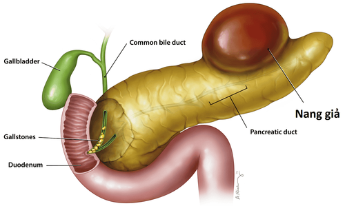
Cystic lesions of the pancreas are associated with a variety of pathologies including neoplastic and noncancerous lesions. The diagnosis and differentiation of these cystic lesions is considered an important issue in planning management.
There are major challenges with diagnostic models for various types of pancreatic cystic lesions. In the era of advanced medicine, methods such as: Ultrasound-guided fine-needle aspiration cytology with chemical and molecular analysis of cyst fluid; EUS-guided fine-needle confocal laser endoscopy (endoscopic ultrasound), via microneedle biopsy; Cholangioscopy, pancreatoscopy... are evaluated as promising in the diagnosis of pancreatic cysts.
2. Prospective studies in the diagnosis of pancreatic cysts
Testing for pancreatic malignancies related mutations increases the diagnostic accuracy of pancreatic cysts. Routine molecular analysis of KRAS/GNAS mutations gives high sensitivity and specificity, reaching 90% in detecting mucosal lesions. However, KRAS mutations still do not grade malignancy. Thus, Springer et al. concluded in their study that mutations in the von Hippel-Lindau (VHL) gene (3p35) with loss of heterozygosity at the genomic position on chromosome 3 and aneuploidy chromosome 3p, is detected in approximately 67% of serous thyroid nodules.
A study performed by Arner et al. mentioned that: “The addition of molecular DNA analysis alters the clinical management of pancreatic cystic lesions, most often when CEA levels are moderate average (45-800 ng/mL) or in the absence of CEA concentrations”. GNAS mutations alone can be detected in up to 66% of mucosal neoplasms, their detection reaching up to 96% of cases when combined with KRAS mutations. They were also found in 100% of IPMN cases with a sensitivity and specificity of 89% and 100%, respectively, for detecting mucocutaneous cysts.
Other mutations investigated include TP53 mutation, deletion mutation in p16/CDKN2A, SMAD4, mutation in TP53, PIK3CA and/or PTEN. They are very sensitive to IPMN. According to a systematic review by Zhang and Pitman, although molecular testing is not a substitute for cytological and chemical testing, it can still add value to diagnostic results.
3. Studies using DNA analysis of follicular fluid appear promising but need further attention
The final randomized clinical study discussed radiographic and imaging modalities of pancreatic cystic lesions. However, further studies are still needed to improve the diagnosis and define a clear strategy for the use of new endoscopic techniques in the diagnosis of pancreatic cysts.
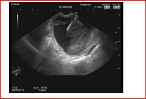
Table of final randomized clinical studies on radiographic and imaging modalities of pancreatic cystic lesions:
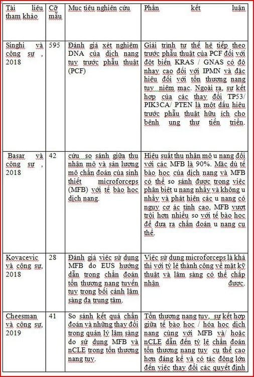
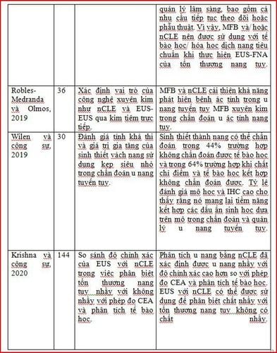
Abbreviation: IPMN: Intracellular papillary mucinous carcinoma; PCL: Injury to pancreatic cyst; EUS: Endoscopic Ultrasound; FNA: Fine Needle Aspiration; NR: Not reported; nCLE: Needle-based confocal laser endoscopy; IHC: Immunohistochemistry. In summary, the real clinical challenge in the management of pancreatic cystic lesions is to determine which patients should undergo pancreatectomy because the cancer has not metastasized early, and which patients with precancerous cystic lesions are at risk. high or the patient has limited/no predisposition to malignancy. By combining the use of EUS modalities with cystic tumor markers, the analysis creates a new diagnostic model for pancreatic cystic lesions. In addition, it really emphasizes that the accurate diagnosis of pancreatic cystic lesions requires a multidisciplinary and multimodal team approach, along with the integration of clinical findings, imaging , cytology, cyst fluid analysis and molecular analysis.
Please dial HOTLINE for more information or register for an appointment HERE. Download MyVinmec app to make appointments faster and to manage your bookings easily.
References Okasha HH, Awad A, El-meligui A, Ezzat R, Aboubakr A, AbouElenin S, El-Husseiny R, Alzamzamy A. Cystic pancreatic lesions, the endless dilemma. World J Gastroenterol 2021; 27(21): 2664-2680 [DOI: 10.3748/wjg.v27.i21.2664]


