This is an automatically translated article.
The article was professionally consulted by Specialist Doctor I Vo Cong Hien - Radiologist - Radiology Department - Vinmec Nha Trang International General Hospital.An intravenous urogram, also known as an intravenous pyelogram (IVP), is an imaging method that helps to examine abnormalities of the urinary system such as the kidneys, ureters, bladder, and urethra.
1. What is an intravenous urology X-ray?
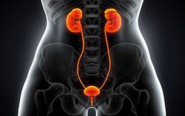
The anatomy of the urinary system in a normal person includes the kidneys (calypses, renal pelvis), ureters, bladder and urethra. When blood flows to the kidneys, it is filtered through structures inside the kidneys to form urine. Then, urine is carried down the bladder through 2 ureters on either side of the body before being discharged through the urethral system.Normally, an abdominal X-ray will not examine these structures except in the case of patients with contrast-enhanced stones in the urinary system. Therefore, to investigate the abnormalities of the urinary system, the doctor will appoint an X-ray of the urinary system by intravenous route or called intravenous nephrography for short.
In this method, contrast dye will be injected into a vein, following the blood to the kidney and being discharged to the outside according to normal physiology. By taking many X-ray films of the urinary system at different times (the feature of the contrast medium is that it does not allow X-rays to pass - creating a white color on the film) it will help to investigate the abnormalities of the urinary system. . This method is X-ray of the urinary system by intravenous line.

Để khảo sát các bất thường của hệ tiết niệu thì bác sĩ sẽ chỉ định chụp X quang hệ tiết niệu bằng đường tĩnh mạch
2. When is an intravenous nephrography indicated?
Intravenous urology x-ray helps to evaluate some of the following abnormalities:Urinary stones: Images of stones in the kidney or in the ureter (contrast or non-contrast) will often present evident on venous radiographs. Repeated urinary tract infections: In case the patient has repeated bladder infections or nephritis, suspected obstruction or urinary tract abnormalities, intravenous urography should be performed. can help identify those risk factors. Hematuria: Blood (or red blood cells) in the urine can be the result of many different conditions such as inflammation, infection or cancer. An intravenous pyelogram can help diagnose or rule out these causes. Urinary tract obstruction or damage: An intravenous x-ray of the urinary system helps doctors diagnose a blockage or injury in the urinary tract.
3. Contraindications of venous nephrography
Intravenous nephrography is contraindicated in the following cases:Renal failure: In order to take radiographs of the urinary system by intravenous route, the most important thing is that the patient's kidneys must be able to filter contrast. Therefore, patients with renal failure are absolutely contraindicated for this method; Allergy/history of anaphylaxis to contrast medium; Women who are in pregnancy.

Chụp X quang hệ tiết niệu bằng đường tĩnh mạch giúp phát hiện nhiễm trùng tiết niệu
4. What preparation should be done before venous nephrography?
Steps to prepare when taking an intravenous pyelogram:Check kidney function to see if there is impaired kidney function by testing for urea and creatinine in the blood. Assess for possible allergy or history of allergy to iodinated contrast media. Fasting (in some cases) to ensure clear X-ray images by eliminating noise signals from food in the intestinal tract. Laxatives are used (in some cases) for the purpose of making the urinary tract X-ray clearer. The patient needs to sign an agreement to accept the risks that may occur during intravenous urography. If a patient with diabetes mellitus is being treated with metformin, the drug should be discontinued at least 2 days prior to the intravenous angiogram. The reason is that Metformin can combine with contrast media and affect kidney function.
5. Steps to take X-ray of urinary system by intravenous route
Steps to take X-ray of the urinary system by intravenous route:The patient lies comfortably on the examination table Conduct contrast injection into a vein in the back of the patient's hand or arm. Over time, the contrast medium begins to be filtered through the kidneys, flows down the ureters, and collects in the bladder. The doctor then takes a series of X-rays of the abdomen, usually taken every 5 to 10 minutes. In particular, before taking the last X-ray of the urinary system, the patient needs to urinate completely. The entire intravenous nephrography procedure usually takes about 30-60 minutes. In some cases, some pictures may be taken several hours after the injection.

Dị ứng với các chất cản quang với các biểu hiện như phát ban ở da là tác dụng phụ có thể gặp
6. Some side effects of intravenous nephrography
Some side effects of intravenous nephrography include:Patients often feel hot in the body, especially at the injection site of contrast dye or sometimes have a metallic taste in the mouth. However, these signs are usually transient and disappear quickly. Allergy to contrast material (rare) with manifestations such as skin rash, itching or mild swelling of the lips. Acute renal failure can also occur, although the incidence is very low. Currently, intravenous urography is not routinely indicated as in the past because of the development of less invasive techniques such as ultrasound, CT and MRI of the urinary system.
Vinmec International General Hospital with a system of modern facilities, medical equipment and a team of experts and doctors with many years of experience in medical examination and treatment, patients can rest assured to visit. examination and treatment at the Hospital.
SEE MORE
What you need to know about X-rays of the urinary system Intravenous X-rays of the urinary system The role of tomography in the diagnosis and treatment of ureteral stones


