This is an automatically translated article.
The article is professionally consulted by Master, Doctor Nguyen Quang Duc - Doctor of Nuclear Medicine - Department of Diagnostic Imaging and Nuclear Medicine - Vinmec Times City International General HospitalGallbladder scintigraphy is an imaging procedure using nuclear scans to evaluate biliary tract disease and gallbladder disease. Gallbladder scintigraphy can detect obstruction of the bile ducts from the liver to the gallbladder and small intestine.
1. Gallbladder scintigraphy
Before cholecystography, the patient needs to fast for about 8 hours. A radioactive tracer will be injected into a vein (usually a vein in the arm). This radioactive tracer is essentially a technetium-labelled iminodiacetic acid compound. After taking into the body, this substance will be metabolized in the liver, excreted in the gallbladder through the bile duct system in and out of the liver. Finally, the radioactive tracer is secreted into the first part of the small intestine. A specialized camera will record images of the tracer moving from the liver to the bile ducts, gallbladder and small intestine.
During the gallbladder scan, the patient needs to remove objects such as jewelry, metal carried around because it can affect the final result.
The steps to take a gallbladder scan include:
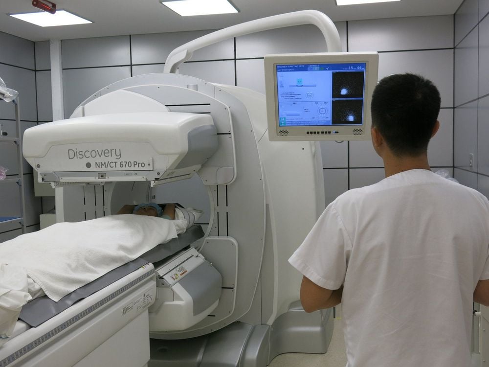
Disinfect the patient's hand - the place where the radioactive tracer was injected. A small amount of radioactive tracer is injected into a vein. The patient lies supine on the table, a large scanning camera is placed just above the patient's abdomen. After the radioactive tracer is injected, the camera scans for the radiation emitted by the tracer. The camera does not produce any radiation. The camera will produce images as the radioactive material passes through the liver and into the gallbladder and small intestine. The first pictures are taken immediately after the tracer injection, can be taken continuously, like a video, or can be taken once for up to an hour and a half after starting. Each radiation scan takes only a few minutes, the patient needs to lie still during each scan so that the image does not become blurred.

2. What does a gallbladder scan mean?
In acute cholecystitis (often due to stones), the gallbladder will not show up on radiographs because radioactive material cannot get inside the gallbladder. This procedure provides a fairly accurate diagnosis (with the exception of false positives in some critically ill patients). However, in clinical practice, scintigraphy of the gallbladder is rarely needed to diagnose acute cholecystitis.
If your doctor suspects non-stoner or obstructive cholecystitis, your doctor will order cholecystography before and after cholecystokinin (stimulating gallbladder contractions). Decreased concentration of radioactivity will guide decreased gallbladder secretion. Decreased gallbladder emptying (measured by excretion rate) would suggest non-stoner or obstructive cholecystitis.
Gallbladder scintigraphy also helps to detect bile leaks and anatomical abnormalities ( common bile duct cysts ). After cholecystectomy, scintigraphy of the gallbladder can help quantify bile flow, identifying dysfunction of the sphincter of Oddi.
Gallbladder scan helps to find the cause of upper and right abdominal pain, check gallbladder function, find the cause of jaundice and find blockage of the ducts from the liver to the gallbladder and small intestine .

3. How long does a gallbladder scan take?
A gallbladder scan usually only takes about 1 to 2 hours, depending on the results it can be done more than a day later. If the patient needs to come back for another gallbladder scan, do not eat any fatty foods before the gallbladder scan.
The patient may not feel anything even when the tracer is injected, or may feel a sting, but most gallbladder procedures are usually painless, try to relax by breathing slowly and deeply.
Patients may experience nausea or abdominal pain when gallbladder stimulants are used during the procedure.
4. Risks when performing gallbladder scan
Allergy to radioactive tracer is very rare as most of these substances will leave the body through urine or feces within 1 day. The toilet should be flushed immediately after each use, but the amount of radiation is so small that there is no risk of harm from post-procedure exposure. Some patients may experience pain or swelling at the injection site after the injection, which can often be relieved by placing a warm, moist compress on the arm. There is a small risk of damage to cells or tissues from radiation, but the potential for harm is usually very low compared to the benefits of this procedure. In cases where scintigraphy cannot be performed or the assessment of biliary tract disease is not useful, such as:
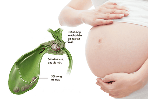
Pregnant patient: Gallbladder scan is not usually indicated during pregnancy because radiation can damage hurt the baby. The patient has recently had a test that involves the use of barium or a medication containing bismuth . The patient cannot lie still during the procedure. The patient is allergic to morphine.

5. Read the results of a gallbladder scan
5.1. Normal results The radioactive tracer flows evenly through the liver, then into the gallbladder and into the first part of the duodenum. The obtained gallbladder scintigraphy was of normal size, shape, and position. 5.2. Abnormal results Tracker is not removed from the blood by the liver: this is a sign of liver disease. The gallbladder does not contract or is empty. Tracker does not reach the gallbladder: signs suggest swelling or ducts blocked by gallstones. The tracer does not reach the first part of the duodenum: This means there is a bile duct blocked by a stone or there may be a tumor, infection, or swelling of the pancreas. Pain occurs when the gallbladder empties. Gallbladder scintigraphy is a technique that needs to be performed in modern medical facilities with good and experienced doctors and technicians. Currently, the Gallbladder Scanning technique is being applied by Vinmec International General Hospital in the diagnosis of biliary tract diseases. Vinmec uses the most modern SPECT/CT Discovery NM/CT 670 Pro system with the most modern 16-series CT in Southeast Asia, manufactured by the world's leading medical equipment company GE Healthcare (USA). The team of doctors and technicians at Vinmec are experienced, well-trained at home and abroad, bringing maximum efficiency to the imaging process.
Please dial HOTLINE for more information or register for an appointment HERE. Download MyVinmec app to make appointments faster and to manage your bookings easily.


