This is an automatically translated article.
The article is professionally consulted by Master, Doctor Pham Thi Thuy Nhung - Dean and Master, Doctor Do Thi Hoang Ha - Laboratory Department - Vinmec Hai Phong International General Hospital.Manual urinalysis is an important subclinical technique, performed for the purpose of detecting, diagnosing and treating a number of diseases affecting human health. change for the best treatment effect.
1. Urine sediment
Urine is one of the very important secretions for human physiology because it contains most of the waste products in the body called urine sediment, which is related to most of the chemical components of the body. body. Therefore, when there are any metabolic changes, be it diseases or metabolic disorders, it also affects the results of urinalysis.2. Manual urine sediment test
Urine sedimentoscopy test is a subclinical test that serves a lot in the detection, diagnosis as well as treatment, monitoring and treatment of diseases, thereby preventing unwanted complications. want to happen.
Test urine sediment by manual method with the aim of finding the tangible elements present in the urine such as red blood cells, white blood cells, epithelial cells, crystals, cylinders..., from which to evaluate assess the patient's condition. Urine microscopy is a manual urine sediment test, which is performed by examining the urine sediment under a microscope with a 10X objective.
3. Urine sediment screening process
To conduct a fresh smear of urine, it is necessary to prepare a few things:
Performer: Staff of the Department of Biochemistry / Hematology Pipetman, slide, optical microscope. Explain to the patient about the test to be done, instruct the patient to collect urine in the correct procedure, in the right way, preferably in the early morning upon waking up. Specify patient information such as name, age, gender, department, room... on the test paper and pay attention to specifying the test order. Perform a fresh smear of urine sediment according to the following steps:
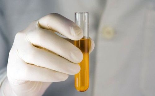
After giving the patient a midstream urine collection in the morning or at any time of the day into the test tube, let the sediment settle after about 1 hour, do not centrifugal. Empty the upper part of the urine, gently shake the residue in the test tube, and remove about 1 drop of the remaining urine residue on a clean slide. Directly examine the prepared slide under the 10X objective. Distinguish between components in urine such as cells, cylinders, and crystals. Determine the level of pathological cells present in the specimen. Record information about important pathological cells on test sheets and store them in the medical facility's data management software. Some results that can be identified after performing a urine sediment test by the fresh endoscopy method are:
Normal: There are few or no white blood cells and red blood cells in the urine, specifically 0- 3 white blood cells/microfield and 0-2 red blood cells/microfield. Flat cells are seen because the cells lining the ureter are degenerated and a small amount of sperm can be seen on urinalysis in men.

Hình ảnh các tế bào trong nước tiểu
Hematuria:
The image of red blood cells in the urine sediment, the naked eye will see red urine, red, small, blue disc-shaped red blood cells. The size of red blood cells can be 8μ if normal and 5-6μ if atrophied, 9-10μ when red blood cells are enlarged. The number of red blood cells can be from 3-20 erythrocytes/microsphere or sometimes > 20 erythrocytes/microfield. Some cases are said to be gross hematuria when we see a large amount of hematuria, when fresh, the urine is pink like meat wash or red, so it takes a while for red blood cells to settle down. under. On the microscopic slide, the urine residue is microscopic densely packed red blood cells. Urinalysis will not show red blood cells in the case of red blood cells that are lysed. Visceral microscopic findings that show red blood cells in the urine or are often suggestive of conditions such as glomerulonephritis, renal tuberculosis, urolithiasis, cystitis, bladder cancer, hematopoietic system disease , blood clotting disorders ... There are also cases where the cause of hematuria is not found. Leukuria:
Fresh examination shows the image of urine leukocytes, also known as urine leukocytes, disc-shaped cells, bright, with granules inside the cells, intact or atrophied or concentrated as a cloud of pus . Normally, white blood cells are 1.5-2 times larger than red blood cells. The white blood cell count may be 3-20 or >20 leukocytes/microfield. If the white blood cell count is >10-20 leukocytes/microsphere or >20 white blood cells/microsphere, it is usually a urinary tract infection. In special cases, > 30 leukocytes/microfield, the image of white blood cells will be dense, with many degenerated white blood cells called pyuria, common in acute and chronic pyelonephritis. Cylindrical urine:
Fresh examination shows the image of cells with a cylindrical structure in the urine, which is essentially mucoprotein, which is a sign of damage to the renal tubules and glomeruli. Cylindrical cylinders can be seen as:
Inner cast (long shape, round head, ringless, transparent) when fever, heavy labor, nephritis... Wax pillar (shorter, bigger than inner cylinder) , iridescent, grey, cracked). Fat casts (luminescent, yellow, clear margin, grooved, rounded tip) are seen in nephrotic syndrome. Or the image of cytoskeleton such as: Granule casts (with large, sweet yellow, round heads) in acute and chronic glomerulonephritis, acute renal failure. RBC casts (agglutinated red blood cells, irregular jagged edges) are seen in glomerulonephritis. Leukocyte casts (leukocytes form, break into short segments) are seen in acute and chronic pyelonephritis. Epithelial column (contains epithelial cells, bright yellow). Bacterial casts If a fresh examination of the urine sediments shows protein and even cylindrical casts, the kidney may have been severely damaged, or vice versa, the prognosis may be better than before. Some signs to note to diagnose the disease when doing urine sediment test are as follows:
Proteinuria + erythrocytes + leukocytes + erythrocyte casts + granulomatous casts + fat casts suggest glomerulonephritis acute. The image of erythrocytes + erythrocyte casts + transparent fibrin indicates bleeding inside the kidney. The image of leukocytes + degenerated white blood cells can diagnose pyelonephritis and pyelonephritis. In addition, other components encountered when testing urine sediment by this method are crystal deposits:
Calcium oxalate in envelope shape, green peanut, with square Uric Acid optical extractor, diamond, block,... yellow, reddish brown. Phosphate rectangular, fern leaf, optically refractive, colorless... Urate spherical, yellow, refractive...

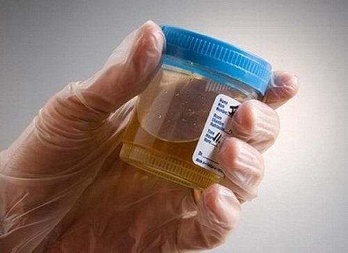
Xét nghiệm cặn nước tiểu bằng phương pháp thủ công là kỹ thuật cận lâm sàng quan trọng
A few crystals that are not commonly encountered in urinalysis by urinalysis such as stellate phosphate dicalcium, bunch of slides and colorless, spherical calcium carbonate, small particles, in pairs, calcium sulphate needle long, prismatic, flattened, colorless. Or amorphous inorganic urine sediments are not a sign of pathology, such as amorphous phosphates, small particles, without a specific shape, dispersed and capable of soluble in acids, amorphous urate. Some inorganic urine sediments show pathology, such as spherical Leucin, with concentric, radial lines, the size of urine Leukocytes and Tyrosin long needle-shaped, bundle wall, colorless or yellow seen in pathology. severe liver stage...
Some cells of medium and large size can be seen on scintioscopy, valuable in diagnosis if observed in large numbers, often a sign of muscle disease Genital organs such as vaginitis, urethritis, cystitis or urinary tract disease, nephrotic syndrome, including:
Urethral epithelial cells are polygonal in shape and contain nuclei. Urethral epithelial cells are large, polygonal, oval, with nuclei Bladder epithelial cells are large, rhombus-shaped, with nuclei Some other cells such as pyelonephritis, renal cells, nuclear carcinoma cells monster in kidney-urinary cancer pathology. Manual urine sediment test is a urine testing technique that requires high accuracy, so it should be performed by highly qualified and experienced laboratory technicians. The results of the urine sediment test are very valuable in the diagnosis of urinary tract diseases as well as systemic diseases and diseases in other organs.
To register for examination and treatment at Vinmec International General Hospital, you can contact Vinmec Health System nationwide, or register online HERE.
MORE:
Meaning of SG in a urine test Time to take a urine test during pregnancy Meaning of KET (Ketone) in urine