This is an automatically translated article.
Posted by Dr Kharitonchyk Aksana - Obstetrician and Gynecologist - Women's Health Center - Vinmec Times City International General HospitalPostpartum is the period to return to normal anatomical and physiological sex organs (except for the mammary glands, which continue to develop to produce milk). This is 6 weeks (42 days) from birth or right after the third stage of labor ends.
1. Changes in mammary glands during pregnancy and lactation
First, let's take a look at the basic features of the breast through each period:
At birth, the mammary glands are not fully developed. It only grows a little while in the womb, but grows a lot during puberty and during pregnancy. During these periods, the breasts are very sensitive, especially during puberty, the mammary glands develop the most.
During puberty, breasts begin to grow when breast cells, which are already lying dormant, receive signals from hormones that tell them it's time to start working. So these cells begin to grow and divide rapidly. Their job is to form the characteristic duct system in the adult breast.
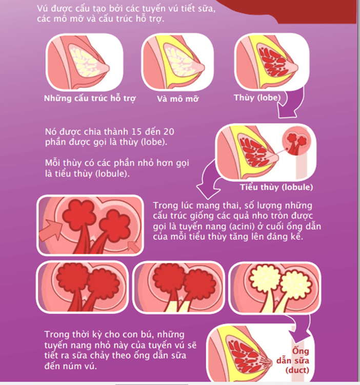
During pregnancy, the breast undergoes another transformation. Only at this point are the breasts truly fully developed and able to produce milk in the lobules, which then transport the milk through the ducts to the nipple.
After breastfeeding, the breasts go through other changes called contractions, and shrink. The cycle of proliferation, milk production and contraction occurs with each pregnancy and it is one of the very important features of the mammary gland.
In adult women, the mammary glands work according to the menstrual cycle and are controlled by the hormones of the hypothalamus - pituitary gland - ovaries. Mammary glands have obvious changes with the menstrual cycle, during pregnancy and lactation that a woman can feel for herself.
2. Breast changes during pregnancy
There is significant growth of ducts, lobules and follicles, due to the effects of luteal and placental sex steroids, hPL, Prolactin...
Development of the mammary glands :
When During pregnancy, the mammary gland achieves complete development, the mammary gland parenchyma proliferates, the epithelial buds transform into lobules, secretory columnar cells are surrounded by a layer of muscle-epithelial cells. Milk ducts are long and branched, and blood vessels proliferate.
During the first 3-4 weeks of pregnancy, the ducts grow significantly and form branches and lobes under the influence of estrogen. By weeks 5-8, the breast is markedly enlarged, with dilation of the superficial veins forming the Haller system, there is a feeling of heaviness and the nipple-areola area becomes darker, Montgomery's granules are prominent due to gland enlargement. residue.
In the second trimester, under the action of progesterone, the formation of lobules has outstripped the development of ducts, colostrum containing fat-free colostrum is formed under the action of Prolactin. From the second half of pregnancy onwards, the breast increases in size due not only to proliferation of mammary epithelium but also to dilation of colostrum-containing follicles, as well as to hypertrophy of epithelial, connective tissue and tissue cells. fat. If this process is stopped by premature delivery, there may be lactation from 16 weeks onwards.
3. Milk production and lactation mechanism:
During pregnancy, the mother's breasts are ready to produce milk. In the second trimester of pregnancy, the mammary glands begin to make colostrum. This milk is yellow, sticky, rich in nutrients and antibodies. After the baby is born and the placenta has been removed, your body begins to produce more milk. Over the next few days, the amount of milk continued to increase and the milk turned white, looking thinner. The secretion and excretion of milk involve many hormones, in which the role of 4 main hormones is estrogen, progesterone, Prolactin and Oxytocin.
Estrogen and Progesterone: help breasts develop, ready for milk production.
Released by the placenta during pregnancy. Estrogen increases the size and number of milk ducts while progesterone helps develop follicles and lobules. High levels of estrogen and progesterone inhibit milk production while the baby is in the womb. When the baby is born and the placenta has shed, the levels of these hormones automatically drop, letting the body know it's time to make milk. Breastfeeding mothers should not take birth control pills containing estrogen because this hormone supplement can reduce breast milk supply.
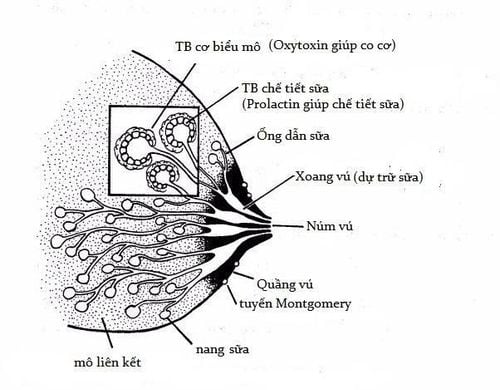
Prolactin: hormone from the anterior pituitary gland, which helps produce milk. During pregnancy, prolactin is also released a lot and has the effect of stimulating the growth of the glandular epithelium. Prolactin levels increase gradually during the first half of pregnancy, from 6 to 9 months, blood levels of Prolactin are 3 times higher than normal, and the mammary epithelium begins to synthesize proteins.
After the baby is born, the level of Prolactin increases.
Breast milk is produced by the physiology of lactation (the Prolactin reflex): when a baby sucks, afferent impulses from the nipple stimulate secretion of Prolactin from the superior pituitary gland. Prolactin acts on the epithelial cells of the milk-producing follicles to stimulate lactation. At this time, milk will be produced and stored in the milk follicles.
If Prolactin levels are too low, breast milk supply will decrease. Therefore, mothers need to breastfeed or pump milk immediately after birth and at regular intervals thereafter.
Prolactin is more active and more milk is produced when the mother's breasts are exhausted after each feeding, when the baby suckles early, sucks often, when the mother expresses milk or feeds the baby at night. Prolactin production is limited when the baby eats other nutrients before breastfeeding, when the baby is placed in the wrong feeding position, when the breast is sore, when the mother is tired and stressed. Oxytocin: secreted from the posterior pituitary gland, released when the baby (or breast pump) begins to suck and pull the nipple into the mouth.
It contracts the muscles around the follicle, pushing the milk out of the follicle, into the milk ducts and moving to the nipple and then into the baby's mouth.
In addition, Oxytocin has the role of contracting the uterine muscles during and after birth, helping this organ shrink to its original size, limiting postpartum bleeding.
4. How does the let-down reflex happen?
Letting go or lactation is a conditioned reflex, pushing milk from the follicles through the ducts to the milk sinuses and nipples. Lactation occurs after 3 to 4 days in calves and 2-3 days in calves. Lactation is caused by a sudden increase in the concentration of Prolactin in the blood, which leads to the synthesis of more milk and the release of milk from the breasts by Oxytocin. This reflex begins a few seconds to a few minutes after the mother begins to breastfeed.
You feel a tingle or slight discomfort in your chest, but you may not feel anything at all. This reflex can also occur at other times, like when a mother hears her baby cry or when she just thinks about her baby.

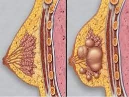
5. What is a breast abscess? Causes, risk factors, symptoms and treatment? Cause-Risk Factors
Breast abscess is a necrotizing, purulent inflammatory condition that forms an abscess of the mammary gland parenchyma or extramammary gland tissue. Most breast abscesses occur after untreated mastitis. 5-11% of nursing mothers with mastitis will develop a breast abscess.
Risk factors for breast abscess include: first pregnancy after age 30, pregnancy over 41 weeks. Postpartum mastitis ineffective treatment, causes of mammary gland blockage, immunocompromised conditions
The most common cause of breast abscess is Staphylococcus aureus that enters the breast tissue through the milk ducts and nipple wounds.
Symptoms
In the early stages, an abscess tends to be limited to one part of the breast. The pathogenesis is similar to that of acute inflammation elsewhere in the body. Loose breast tissue combined with milk stagnation creates a favorable environment for bacteria to grow and quickly disperse into the tissues through the milk ducts.
Mothers often have breast abscess in two stages:
The first month after giving birth for the first time, because breastfeeding experience and inadequate breast hygiene can lead to the risk of nipple damage. The weaning phase, which leads to stagnation of milk. In addition, after 6 months, babies develop teeth that can also hurt the nipples. Symptoms of a breast abscess include:
A palpable, mobile mass in the diseased breast. However, in some cases, the abscess is located deep in the breast tissue and cannot be palpable. The diseased breast side is swollen Hot, red, painful in the abscess area Fever, fatigue Ipsilateral axillary lymph nodes Subclinical:
Blood count: Bach Elevated platelet count, elevated CRP, pus culture for bacteria Ultrasound has an empty or mixed echogenic mass in the mammary gland tissue
6. Treatment of breast abscess
General management
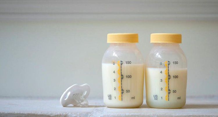
Cold application. Continue breastfeeding or express milk by machine or by hand (Cut milk in case of non-breastfeeding or severe infection, recurrent abscess).
Antibiotics - anti-inflammatory - pain relief. Principles of antibiotic treatment:
Antibiotic treatment as soon as a confirmed diagnosis is made. Adjust antibiotics when the results of the antibiogram are available. Antibiotic treatment before and 10-14 days after drainage of pus. Surgical treatment
Depending on the size of the abscess and the experience of the clinician, the clinician will decide to aspirate the abscess or drain the abscess.
Vinmec International General Hospital is a high-quality medical facility in Vietnam with a team of highly qualified medical professionals, well-trained, domestic and foreign, and experienced.
A system of modern and advanced medical equipment, possessing many of the best machines in the world, helping to detect many difficult and dangerous diseases in a short time, supporting the diagnosis and treatment of doctors the most effective. The hospital space is designed according to 5-star hotel standards, giving patients comfort, friendliness and peace of mind.
Please dial HOTLINE for more information or register for an appointment HERE. Download MyVinmec app to make appointments faster and to manage your bookings easily.


