This is an automatically translated article.
The article is professionally consulted by Dr. Pham Quoc Thanh - Radiologist - Department of Diagnostic Imaging - Vinmec Hai Phong International General Hospital.Magnetic resonance angiography with non-specific contrast injection is a special method used to detect diseases that are directly related to the lymphatic vessels and lymph nodes.
1. What is Magnetic Resonance Imaging with Magnetic Contrast?
Magnetic resonance lymphatic system has been used since the 1990s. Magnetic resonance imaging has many advantages over lymphatic scintigraphy such as high spatial and temporal resolution, 3-D, exposure-free imaging. radiation like other conventional imaging methods. Magnetic resonance imaging in medicine is also abbreviated as MRI.In addition to the advent of magnetic contrast agents and new techniques in magnetic resonance imaging, magnetic resonance imaging allows the evaluation of lymphatic vessels and lymph nodes.
Lymphatic resonance with specific contrast agents such as ultrasmall iron oxide particles (USPIO: Ultrasmall iron oxide particles) is a non-invasive method that allows the evaluation and analysis of the lymphatic system after injection of the substance. intravenous contrast agent. Routine imaging of lymph nodes to assess size and morphology can lead to confusion between benign and malignant lymph nodes. The normal lymph nodes will absorb the iron ions in the contrast agent, shortening the duration of T2-weighted images, resulting in loss of T2-weighted signal after 24 h. It is therefore possible to distinguish metastatic lymph nodes regardless of size criteria because reticuloendothelial cells are replaced by metastatic cells. Metastatic cells do not absorb USPIO, so there is no signal loss on T2-weighted images. USPIO is therefore called a negative magnetic contrast agent. MRI with USPIO injection allows detection of small metastases.

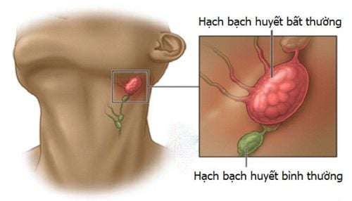
Chụp cộng hưởng từ bạch mạch cho phép đánh giá được mạch máu hạch bạch huyết
2. Difference between lymphatic magnetic resonance with injection of non-specific contrast and with injection of specific contrast agent.
Essentially, these two methods of resonance imaging are similar in terms of effectiveness and imaging procedures. The only difference between the two methods of injection is in the method of injection and the purpose of evaluation.Magnetic resonance angiography with injection of specific contrast agent
Specific contrast agent such as ultrasmall iron oxide particles (USPIO: Ultrasmall iron oxide particles ) Intravenous administration Evaluation of lymph nodes for size , morphology can identify benign and malignant lymph nodes Lymphatic resonance imaging with injection of non-specific magnetic contrast
Conventional contrast contrast injection into the interstitial tissue Structural assessment anatomy as well as part of the lymphatic system function. Engraftment of the lymphatic system (including blood vessels and lymph nodes) allows drainage pathways and sentinel lymph nodes to be identified as targets for biopsy. In addition, it also has the ability to provide information about the functional status of lymphatic transport to the lymphatic vessels and lymph nodes in the injured limb.
3. Indications and contraindications for lymphatic magnetic resonance imaging with injection of specific magnetic contrast agents
Indications:When lesions of the lymphatic system are suspected. Absolute contraindications i
Patients wear electronic devices such as: pacemakers, defibrillators, cochlear implants, automatic subcutaneous drug injection devices, Neurostimulator... Surgical forceps with intracranial metal, eye socket, blood vessels within 6 months. Severe patients need resuscitation equipment next to the body Relative contraindications
Metal surgical forceps >6 months Patient is afraid of the dark or alone
4 . Steps to conduct lymphatic magnetic resonance imaging with injection of specific magnetic contrast agent
Before taking the scanPatients need to master the following requirements:
No need to fast. The patient is carefully explained about the procedure to cooperate well with the doctor. Check for contraindications Instruct the patient to change into the MR dressing room and remove contraindicated items. Have a copy of the request from a clinician with a clear diagnosis or a complete medical record (if necessary) Patient position
Place the patient on the examination table, place the patient's feet towards the machine frame, Use a suitable receiver coil. Technicians can observe and communicate directly with the patient from the control room. Technique:
Intravenous injection of contrast agent from USPIO Injection dose: 2.6mg/kg Survey procedure: Cross-sectional T1WGRE, Cross-sectional T2WFSE, Cross-sectional T2WGRE before and 24 hours after injection, 3DT1WGRE. Slice thickness: 3mm Cross-sectional: Mostly cross-sectional Combination of other sections depending on the survey area, for example: Axillary assessment for breast cancer: need Oblique Sagittal view Pelvic: Horizontal oblique ( obturator plane) Verification of the results:
Comparison of pre- and post-injection images Metastatic lymph nodes do not absorb USPIO so there is no loss of signal on T2W. Some patterns of USPIO uptake are as follows: Malignant lesions do not capture USPIO at all, drug uptake is heterogeneous, focal defects are scattered, and peripheral signal loss remains central. Lymph node signal heterogeneity, irregular margins, and central necrosis of lymph nodes suggest malignancy. However, this result excludes malignancies < 3 mm and outside the study area. USPIO shortens the duration of T1W, thus increasing the signal on T1W. Lymphatic vessels are one of the main components of the immune system, helping to fight the invasion of harmful bacteria, metastatic cells. Therefore, people need to take good care of their lymphatic vessels by seeing a doctor when there are suspicious signs.

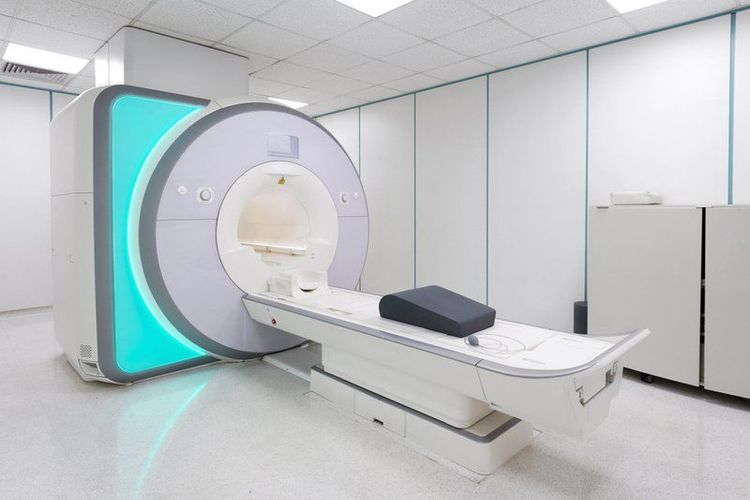
Máy chụp cộng hưởng từ 3.0 Tesla cộng nghệ Silent
Silent technology is especially beneficial for patients who are children, the elderly, weak health patients and patients undergoing surgery. Limiting noise, creating comfort and reducing stress for customers during the shooting process, helping to capture better quality images and shorten the shooting time. Magnetic resonance imaging technology is the technology applied in the most popular and safest imaging method today because of its accuracy, non-invasiveness and non-X-ray use. The circuit will not cause accidents, affecting the patient's health and customers need to make an appointment in advance so that the hospital can prepare medicine during the examination and do not have to wait. All health checks at Vinmec are always performed by a team of highly qualified doctors with many years of experience in the profession.
Master. Doctor. Pham Quoc Thanh has received intensive training and participated in many national and international scientific conferences on diagnostic imaging. The doctor has 13 years of experience in the field of diagnostic imaging and is currently a doctor at the Department of Diagnostic Imaging, Vinmec Hai Phong International General Hospital.
To register for an examination at Vinmec International General Hospital, you can contact the nationwide Vinmec Health System Hotline, or register online HERE.


