This is an automatically translated article.
The article is made by Master, Doctor Ton Nu Tra My - Department of Diagnostic Imaging - Vinmec Central Park International General Hospital.
Magnetic resonance imaging (MRI) of the orbit and optic nerve with injection of magnetic contrast is a modern imaging technique that produces clear images of the orbit and optic nerve that cannot be detected by other imaging methods. Others cannot help diagnose diseases of the eyeball, posterior eyeball, optic nerve tumor,...
1. Learn about magnetic resonance imaging of the orbit and optic nerve with injection of magnetic contrast?
Damage to the orbit and optic nerve if not diagnosed early and treated promptly can lead to blindness. In which these lesions are all very small details in the skull that are difficult to detect through conventional cranial magnetic resonance or cranial magnetic resonance without injection of magnetic contrast. Therefore, the patient should be assigned a special magnetic resonance technique, which is magnetic resonance imaging of the orbit and optic nerve with injection of magnetic contrast agent. This method uses a magnetic field and radio waves simultaneously using contrast material, thus helping to distinguish between normal and pathological structures 🡪 high diagnostic sensitivity and specificity. This technique is widely applied in the diagnosis of pathology of the eyeball, retrobulbar, retrobulbar tumor, pathology of the oculomotor muscles, lesions in the cone including vascular abnormalities, optic neuroma. Sensory and extra-cone lesions including orbital abscess, facial nerve tumor, bone lesions...

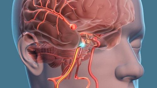
Vị trí dây thần kinh thị giác trên hình ảnh giải phẫu hốc mắt
2. Indications and contraindications
2.1 Indication Cases of magnetic resonance imaging of the orbit and optic nerve without drug injection do not detect lesions but still suspect pathology. Clinically, there are signs of visual loss, clinical examination is consistent with intracranial lesions before or after visual interference. Patients have visual disturbances such as blurred vision, color disturbances, .. but not found. cause by other diagnostic means Suspicion of vascular abnormalities of the orbit such as arteriovenous malformation, carotid-cavernous fistula, .. Eyeball tumor such as retinoblastoma, melanoma, lacrimal gland tumor Abscess mass in the eye Meningiomas of the optic nerve Ophthalmic disease Thyroid glioma optic nerve glioma Follow-up after medical as well as surgical treatment, embolization...
2.2 Contraindications Patients with pacemakers (absolute contraindications) In people with magnetic metals (relative contraindications), especially with metallic foreign bodies inside the eye sockets People with a history of allergy to contrast agents. People who are afraid of enclosed spaces, unable to lie still

Phụ nữ mang thai dưới 12 tuần không nên chụp MRI hốc mắt và thần kinh thị giác
3. Magnetic resonance imaging procedure of the orbit and optic nerve with injection of magnetic contrast agent
3.1 Preparation The patient is clearly explained about the purpose of the scan and the risks of complications There is a commitment to inject contrast from the patient or the patient's family There is a letter of appointment from the clinician. ready with a suspect diagnosis The patient is instructed by the technician to change into the imaging room, remove jewelry, watches, phones, metal objects,... There is enough sedation for patients who cannot lying still or child, contrast agent, anti-anaphylaxis box Steps
Step 1: The patient is moved into the machine compartment. The patient is instructed to lie on his back on the table, then move the table into the magnetic field of the machine
Step 2: The technician performs the imaging technique from the remote control room. The technique includes the following steps:
Localization Selecting the appropriate diagnostic pulse sequences clinically. Performs common pulse sequences: T1, T2, Flair with cutting directions including horizontal, horizontal, and vertical. The transverse direction is along the optic nerve axis, the vertical direction is along the optic nerve axis on each side, the transverse direction is the direction perpendicular to the optic nerve axis. These pulses to the fat removal mode (Fat - sat). Then proceed to run each pulse.
Step 3: Run each pulse and process the acquired images to select images that clearly reveal the pathology
Step 4: Inject contrast. Intravenous contrast injection. Take a T1 pulse to remove fat in 3 directions to cut the space for the orbit (according to the axis is the optic nerve).
Step 5: Read the results from the radiologist.

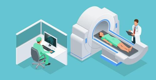
Kết quả chụp cộng hưởng từ giúp bác sĩ tìm nguyên nhân gây bệnh và có phác đồ điều trị tốt nhất
4. Accidents related to technology
Allergic reactions to contrast agents from mild to severe including: nausea, vomiting, urticaria, gastrointestinal disturbances, bronchospasm,... . At worst, anaphylaxis can be fatal, but this case is rare. It is necessary to have a timely treatment attitude on a case-by-case basis, the imaging room needs to have adequate anti-shock equipment. Decreased liver and kidney function. Tell your doctor about your history of kidney disease. Magnetic fields may not be harmful, but they can damage implants or compromise image quality. Vinmec International General Hospital is one of the hospitals that not only ensures professional quality with a team of leading doctors, modern equipment and technology, but also stands out for its examination and consulting services. and comprehensive, professional medical treatment; civilized, polite, safe and sterile medical examination and treatment space. Customers when choosing to perform tests here can be completely assured of the accuracy of test results.
Master. Dr. Ton Nu Tra My is currently a Doctor of Radiology, Vinmec Central Park International General Hospital. Dr. Tra My used to be a lecturer in the Department of Diagnostic Imaging, Hue University of Medicine and Pharmacy and had a long time working at the Department of Diagnostic Imaging, Hue University of Medicine and Pharmacy Hospital.
To register for examination and treatment at Vinmec International General Hospital, customers can call Hotlines of hospitals or register online HERE.