This is an automatically translated article.
The article is expertly consulted by Master, Doctor Trinh Thi Phuong Nga - Radiologist - Department of Diagnostic Imaging and Nuclear Medicine - Vinmec Times City International General Hospital.Magnetic resonance imaging of the lumbar spine uses magnetic waves to create clear, detailed images of the lumbar spine and surrounding tissues. By injecting magnetic contrast, this means better visualization of lesions in this area and less allergic reactions than contrast agents used in computed tomography.
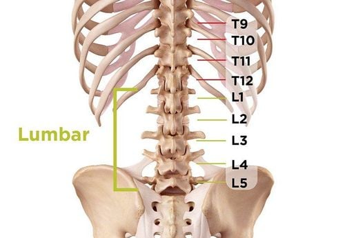
1. What is an MRI of the lumbar spine?
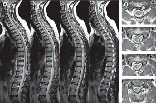
Magnetic resonance imaging (MRI) is a non-invasive technique used to diagnose medical conditions based on the principle of using strong magnetic fields, radio waves and computers to create detailed images. internal body structure. Although no X-ray radiation is used, MRI images are very detailed, allowing doctors to examine the body and detect disease.Currently, MRI is the most sensitive imaging test available for problems in the spine in general and the lumbar spine in particular. The reason is because the anatomical structure in this area is relatively complicated, including many different components, different densities such as bones, discs, ligaments, nerves and surrounding soft tissue that on MRI images can show. better distinguishable.
2. When to take mri of the lumbar spine?
Images in an MRI of the lumbar spine are used to evaluate or detect:Anatomical abnormalities of the spine and alignment.
● Birth defects in the vertebrae or spinal cord.
Injury to bones, discs, ligaments or spinal cord.
Herniated disc and joint pathology. These are common causes of severe lower back pain and sciatica.
Compression or inflammation of the spinal cord and nerves.
Infection of the vertebrae, discs, spinal cord or membranes.
● Tumors in the vertebrae, spinal cord, nerves or surrounding soft tissues.
In addition, an MRI of the lumbar spine with magnetic contrast injection is also used to help plan procedures such as decompression of a pinched nerve, spinal fusion, or steroid injections. In the past, pain-relieving steroid injections were usually performed with X-ray guidance, but since the advent of MRI, the advantage has been to reduce the risk of radiation exposure for patients.
At the same time, MRI of the spine also detects other possible causes of back pain such as bone damage or changes in the spine after surgery, such as scarring or infection.
MRI cột sống cũng phát hiện các nguyên nhân có thể khác gây đau lưng như gãy xương hay những thay đổi ở cột sống sau khi phẫu thuật
3. Magnetic resonance imaging procedure of the lumbar spine with injection of magnetic contrast agent
You will need to wear a hospital gown to remove metal objects on your body, including medical equipment such as hearing aids. In cases where patients cannot remove devices such as pacemakers (with non-demagnetized ones), MRI becomes contraindicated.The patient should be instructed to fast for at least 6 hours due to the injection of magnetic contrast. However, patients still receive medication to treat chronic diseases. In addition, when having contrast injection, you need to declare if you have asthma or are allergic to iodinated contrast agents, drugs, foods or the environment.
Although MRI often uses a contrast agent, Gadolinium, which is less likely to cause an allergic reaction than an iodinated contrast agent, the risk still exists. Also, you may need blood tests to determine your kidney function before taking the contrast agent.
For people who are too nervous, excited, afraid of being closed or uncooperative when taking MRI, especially in infants and young children, low-dose sedation should be indicated.
Once prepared, you will be arranged to lie on a table that slides into the body of a large cylindrical magnet. The technician will control the entire capture process and control the signal into the area to be surveyed.
During shooting, the camera will make noise. You can use earplugs or headphones to listen to your favorite music, avoiding noise for a long time.
The environment in the MRI machine will make some people afraid, called the incubator syndrome. However, you are always observed and monitored by technicians from the outside as well as communicating and asking if necessary.
An initial film called magnetic resonance imaging of the lumbar spine without the injection of magnetic contrast. Then, a dose of contrast material is injected into your body through a vein in your arm. The technician will monitor the time of injection in the area to be taken and repeat the scan, magnetic resonance imaging of the lumbar spine with injection of magnetic contrast medicine.
Once the scan is complete, you can go home and resume your normal activities. If you have used sedatives, you need to be kept under observation until the drug wears off completely and needs to be brought home.
The entire lumbar spine MRI with contrast injection is usually completed within 30 to 60 minutes. However, the analysis of the results will take more time and the results will be returned to the appointing doctor for further diagnosis and treatment.
4. What should be monitored after magnetic resonance imaging of the lumbar spine with injection of magnetic contrast agent?
An MRI scan is completely painless. However, intravenous injection of contrast material to obtain better images can cause discomfort and bruising at the injection site....In addition, there is also a very small chance of The drug causes skin irritation at the injection site, some patients may have a temporary metallic taste in the mouth after the injection of the contrast agent. .
5. The benefits and risks of magnetic resonance imaging of the lumbar spine with contrast injection
5.1 Benefits
MRI is a non-invasive imaging technique that does not involve radiation exposure.MRI images of the spine are clearer and more detailed than those obtained with other imaging methods, especially in diseases of the spinal cord and nerves where abnormalities can be obscured by bone in other imaging methods.
The contrast material in MRI is a contrast agent from Gadolinium, which is less likely to cause allergic reactions than the iodine-based contrast materials used for x-rays and computed tomography.
Furthermore, MRI is also useful for evaluating spinal cord injuries, helping to diagnose or rule out acute spinal cord compression and evaluate ligament injuries, detecting tumors, abscesses, and other soft tissue masses. near the spinal cord.
Finally, MRI is the preferred method for evaluating potential complications of surgery, including bleeding, scarring, infection, and recurrence of herniated discs.

MRI là một kỹ thuật hình ảnh không xâm lấn, không liên quan đến việc phơi nhiễm bức xạ
5.2 Risks
MRI poses virtually no risk to the patient if appropriate safety guidelines are followed.The risks, if any, are often associated with sedation. Therefore, indications and dosages need to be considered in each patient as well as follow-up to limit these risks.
In summary, magnetic resonance imaging of the lumbar spine with injection of magnetic contrast is a useful imaging tool to help investigate local pathologies very well. Compared with contrast-enhanced computed tomography, this means has many outstanding advantages in minimizing the risk of radiation exposure as well as drug allergies.
Vinmec International General Hospital is one of the hospitals that not only ensures professional quality with a team of leading medical doctors, modern equipment and technology, but also stands out for its examination and consultation services. comprehensive and professional medical consultation and treatment; civilized, polite, safe and sterile medical examination and treatment space.
Doctor Trinh Thi Phuong Nga is a radiologist with nearly 20 years of experience in diagnostic imaging. Currently, the doctor is working at - Department of Diagnostic Imaging - Vinmec Times City International General Hospital.
Customers can directly go to Vinmec Health system nationwide to visit or contact the hotline here for support.