This is an automatically translated article.
This article is professionally consulted by Master, Resident Doctor Tran Duc Tuan - Department of Diagnostic Imaging - Vinmec Central Park International General Hospital.MRI is becoming increasingly popular in the investigation of bone and nerve related structures, especially the lumbar spine. This medium provides objective, clear, reconstructive images and is very useful in solving pathological problems in the lumbar spine.
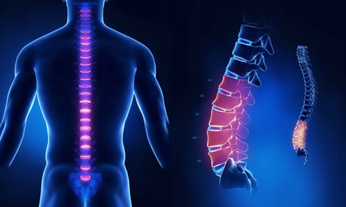
1. What is magnetic resonance imaging (MRI) of the lumbar spine?
Magnetic resonance imaging (MRI) of the lumbar spine is a safe and completely painless test. The principle of magnetic resonance imaging is to use magnetic energy and radio waves to create detailed images of the lumbar spine, including bones, discs, ligaments, spinal cord, nerve roots. and other structures in the lower back.
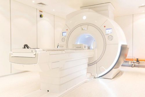
Magnetic resonance imaging differs from computed tomography in that it does not use radiant energy. An MRI scanner consists of a large tubular magnet with a tunnel that is centered on the patient. The patient is placed on a table and slides into the tunnel, inside the magnetic cage. Some centers have MRI machines that have larger openings and become helpful for patients with claustrophobia.
During the test, the radio waves are adjusted to work properly on the areas to be investigated. Multi-dimensional tomographic signal will be computer processed and reconstructed. These images can be reconstructed into a three-dimensional image of the scanned area. This helps to pinpoint problems in the lumbar spine area with suspected abnormalities in this area.
Chụp cộng hưởng từ khác với chụp cắt lớp vi tính là không sử dụng năng lượng bức xạ
2. What is the indication for magnetic resonance imaging of the lumbar spine?
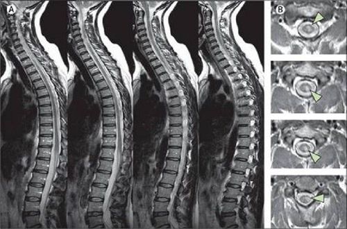
Unlike computed tomography, magnetic resonance imaging of the lumbar spine can detect many different conditions and diseases of the lumbar spine, including problems with the bones (vertebrae), soft tissues (such as the spinal cord), nerves, and discs.MRI is sometimes done to evaluate the anatomical structure of the lumbar spine, to help plan spinal surgery, or to monitor changes in the spine after surgery. For example, magnetic resonance imaging of the lumbar spine can find spinal stenosis, an abnormally narrowed portion of the spinal cord, and surgery is required to clear the narrowing.
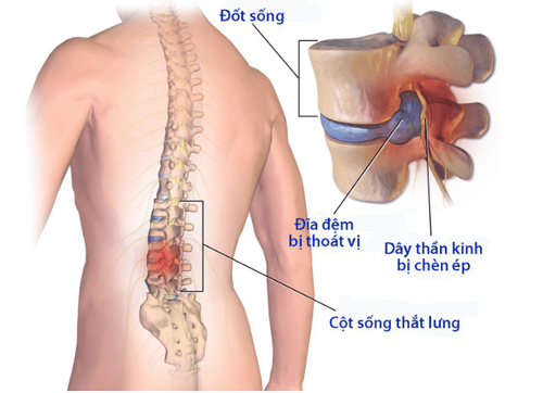
Besides, magnetic resonance imaging of the lumbar spine can also evaluate the discs in terms of structure and position, whether there is a herniated disc inserted into the spinal cord or nerves.
Additionally, an advantage of lumbar spine MRI is that it is helpful in evaluating symptoms such as lower back pain, leg pain, numbness, tingling, or weakness in the limbs as well as problems with bladder control. optics and intestines. At the same time, MRI can also help diagnose tumors, bleeding, swelling, inflammation, infection in the vertebrae or surrounding tissues.
3. Magnetic resonance imaging procedure of the lumbar spine without injection of magnetic contrast agent
Magnetic resonance imaging of the lumbar spine without contrast injection usually does not require any special preparation. However, the technician will ask you to remove any objects on your body that contain metal, such as eyeglasses and jewelry, as these can cause interference in the magnetic environment.These metal objects also include electronic devices such as hearing aids. For non-removable devices such as pacemakers or in patients with artificial joint replacements or metal fixations, these patients are contraindicated for general MRI.
To get the highest quality MRI results, you will need to keep your body still during the scan. For this reason, sedation may be indicated if the patient is very anxious, agitated, or in infants and young children. If this medication is needed, the patient should fast before that to minimize the risk of aspiration. In addition, it is important to inform the MRI technician of any medical conditions, allergies, previous drug reactions, or if you are pregnant.
An MRI of the lumbar spine without injecting contrast usually takes about 30-60 minutes to perform. You will lie still on the scanning table that slides into the machine room while the technician can still communicate verbally with you. If the sound in the device when operating is too noisy, you can use earplugs or wear headphones to your favorite music.
Meanwhile, the magnetic coil will rotate around the body and create an image of the lumbar spine. Each scan will take up to a few minutes until the end of the lumbar area to be examined.
During the procedure, whenever you feel uncomfortable, you can press the button to contact the technician to pause for reassurance. However, you are monitored at all times and will be connected to a machine that monitors your heart rate, breathing rate and oxygen level to ensure you are kept safe at all times.
After the MRI scan is over, the table slides out and the technician will help you get up from the table. If sedation is used, you may be transferred to a recovery area to wait until you are awake before you can leave. Most sedation should go away within 1-2 hours and you can immediately return to your normal activities, routines, and diet.
Chụp cộng hưởng từ cột sống thắt lưng không tiêm thuốc đối quang từ thường không cần bất kỳ sự chuẩn bị đặc biệt
4. What are the results of magnetic resonance imaging of the lumbar spine without injection of magnetic contrast?
The MRI images will be viewed by a specially trained radiologist. The results will be sent back to the doctor who ordered the initial appointment.Common pathologies on magnetic resonance imaging of the lumbar spine are often associated with skeletal structures, discs, ligaments and nerves. Among them, acute neurological diseases are especially important. Thereby, by combining with clinical evidence, doctors will make immediate intervention decisions, in order to minimize the damage to a minimum as well as prevent sequelae.
For chronic musculoskeletal diseases, the results of magnetic resonance imaging of the lumbar spine as a factor contribute to the diagnosis and decision of surgical intervention, depending on the clinical situation with the goal of to the highest benefit, the possibility of recovery for the patient.
In summary, magnetic resonance imaging of the lumbar spine without contrast is an advanced imaging tool for examining structures in this area. The results of the scan not only help diagnose the disease but also guide the treatment, contributing to urgent decisions in critical situations affecting the nerves, preserving the patient's function.
Vinmec International General Hospital is one of the hospitals that not only ensures professional quality with a team of leading medical doctors, modern equipment and technology, but also stands out for its examination and consultation services. comprehensive and professional medical consultation and treatment; civilized, polite, safe and sterile medical examination and treatment space.
Master, Doctor Tran Duc Tuan is well-trained in the country and many centers have prestigious medical background in the world such as: Australia, Singapore, Thailand... The doctor has many years of experience in the field of medicine. imaging and endovascular and extravascular interventions. Currently, the doctor is working at the Department of Diagnostic Imaging and Interventional Vascular Unit - Vinmec Central Park International General Hospital.
Customers can directly go to Vinmec Health system nationwide to visit or contact the hotline here for support.