This is an automatically translated article.
The article is professionally consulted by Dr. Nguyen Dinh Hung - Diagnostic Imaging - Department of Diagnostic Imaging - Vinmec Hai Phong International General Hospital.
Anal fistula is a pathology formed from the abscesses near the anus (accounting for 50%). The cause is usually due to occlusion of the sebaceous glands, long-term causing infection, abscess formation and gradually forming fistula if not diagnosed and treated. Magnetic resonance imaging is an effective diagnostic tool in determining the fistula and the location of deep-seated abscesses.
1. Outline
Anorectal fistulas are small tunnels formed from abscesses near the anus, these fistulas can connect between the rectum and the skin near the anus. When an infected abscess near the anus is formed, the accumulation of pus inside will tend to find its way out, these pus can go into the anastomosis with the rectum, go out to the skin near the anus. anal or both directions.
The typical symptoms of an anorectal fistula are mainly irritation of the perianal skin; pain when sitting, coughing, defecating. In addition, the symptoms that cause patients to complain a lot are fistulas, pus, and fluid when defecating.
Not only causing uncomfortable symptoms for patients, anorectal fistulas are often difficult to treat and easy to recur. The mainstay of treatment is surgery because these fistulas rarely heal on their own. The principle is that it is necessary to identify and evaluate the entire fistula for surgery, so imaging methods such as magnetic resonance are very important in the diagnosis and treatment of anorectal fistula. colon.
Currently, high-field magnetic resonance imaging (High-field MRI) is the best method to provide accurate anatomical path of the anal canal, sphincter and fistula correlation with the ethmoid structure. .
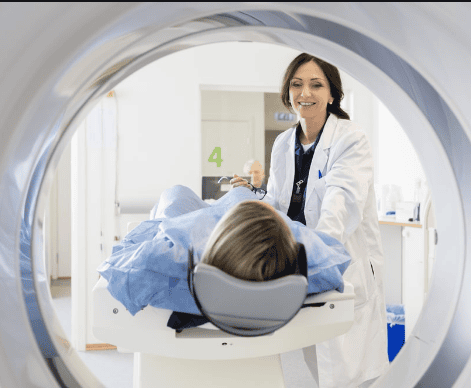
Chụp cộng hưởng từ từ lực cao cung cấp chính xác đường đi giải phẫu của ống hậu môn
2. Indications for magnetic resonance imaging of anorectal fistula
Locate and evaluate primary fistulas or residual abscesses in the anorectal region; ● Locate and evaluate secondary fistulas;
● Correlation of the fistula with pelvic floor structures;
Evaluation of the anal sphincter complex.
3. Contraindications
3.1. Relative contraindications
History of implantation of electromagnetic devices such as pacing blood, defibrillator, cochlear electrode, continuous subcutaneous injection pump; ● History of implantation of metal surgical instruments in the skull, orbit, and blood vessels < 6 months;
● Patient needs critical care equipment nearby.

Bệnh nhân có tiền sử cấy ghép máy tạo nhịp chống chỉ định tương đối thực hiện
3.2. Relative contraindications
Metal surgical facilities > 6 months. ● Fear of the dark, fear of narrow spaces, fear of loneliness.
4. Preparation for anorectal fistula magnetic resonance imaging
4.1. Human
The radiologist is the reader of the results; ● Diagnostic imaging technician;
● Nursing.
4.2. Medical equipment and supplies
Magnetic resonance machines with a force of 1 Tesla or more; ● Medicines: contrast agents, tranquilizers, skin and mucosal antiseptics, shock-absorbing boxes;
● Medical supplies: intravenous needles, syringes, distilled water or physiological saline, gloves, wipes.
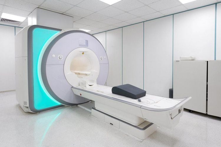
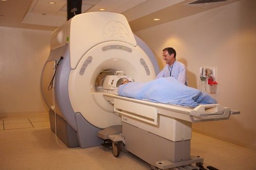
Hình ảnh máy cộng hưởng từ có từ lực tại Vinmec
4.3. Patient
Patients need to be explained and instructed on what to do before conducting magnetic resonance imaging; ● Remove contraindicated items;
● Sedation can be used in patients with anxiety and syndromes such as fear of the dark, fear of narrow spaces, and fear of loneliness.
5. Steps to conduct magnetic resonance imaging of anorectal fistula
5.1. Patient Pose
● Place the patient in the supine position, then select and position the receiver.
● The table is moved into the magnetic field of the blood and locates the area to be scanned.

Bệnh nhân chụp MRi ở từ tế nằm ngửa
5.2. Pulse sequences selected for capture
● Capture the original lesion location;
Non-contrast pulse sequences are in the following order: transverse-oblique plane T1W FSE, transverse-oblique plane T2W FSE or alternatively three-dimensional T2W and STIR pulse sequences;
● Conduct contrast injection from 0.1 mmol Gadolinium/kg body weight, speed 2m/s;
● Take a T1W pulse sequence with fat removal after injecting magnetic contrast.
5.3. Evaluation of shooting results
● The radiograph should clearly show the anorectal wall structures.
● Identify the fistula, path and infiltrates around the fistula and deep abscesses that are not clinically detectable.
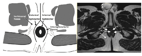
Phim chụp MRI rò hậu môn
6. Complications and how to handle them
● For fearful, agitated patients, comfort and encouragement are needed. If you are too worried, you can use sedation under the supervision of an Anesthesiologist.
Vinmec International General Hospital is currently one of the major hospitals with modern machinery and equipment for general medical examination and treatment procedures and accurate and modern MRI for brain diseases. , cerebral blood vessels in particular.
Especially, Vinmec International General Hospital is the first unit in Southeast Asia to put into use the new 3.0 Tesla Silent Resonance Imaging machine from the US manufacturer GE Healthcare.
The machine currently applies the safest and most accurate magnetic resonance imaging technology available today, without using X-rays, non-invasive. Silent technology is very beneficial for patients who are young children, the elderly, patients with weak health or have just had surgery.
Doctor Nguyen Dinh Hung has over 10 years of experience in the field of diagnostic imaging (Ultrasound, CT, MRI). Trained and practiced on hepatobiliary interventional radiology at Bach Mai Hospital (Intervention under ultrasound guidance, DSA, CT...) and deployed at the Diagnostic Imaging Department of Viet Tiep Hospital Hai Phong. Currently, he is a doctor at the Diagnostic Imaging Department of Vinmec Hai Phong International General Hospital.
To register for an examination at Vinmec International General Hospital, you can contact the nationwide Vinmec Health System Hotline, or register online HERE.


