This is an automatically translated article.
The article was professionally consulted by Specialist Doctor I Nguyen Thanh Hai - Radiologist - Department of Diagnostic Imaging and Nuclear Medicine - Vinmec Times City International General Hospital.Tractography plays an important role in the diagnosis of neurological diseases in general and invasive brain tumors in particular. The method is used to find damage in the nerve fiber bundle or the relationship between the lesion and the nerves to avoid affecting the nerve fiber bundles every time the lesion is intervened.
1. Overview of invasive brain tumors
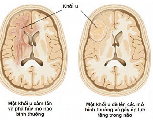
Brain tumors are tumors that form in the skull. There are more than 120 types of brain tumors, but the most common are meningiomas, pituitary tumors, gliomas, and invasive brain tumors that have metastasized to the brain.Brain tumors have 2 types: benign brain tumors and malignant brain tumors:
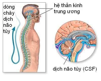
Benign brain tumors tend to grow slowly and do not metastasize to other areas of the brain. Most benign brain tumors can be removed and cured, while others cannot because of the size and location of the tumor. In some cases, benign brain tumors still have the ability to grow and transform into malignant tumors. Malignant brain tumors include tumors in the brain that grow rapidly and invade surrounding healthy brain tissues (cerebrospinal fluid, meninges, spinal cord...).
2. Invasive brain tumor symptoms
The symptoms of a brain tumor vary greatly depending on the type, size, and location of the tumor in the brain.Headache. Weak half man. Vomiting, persistent nausea.

Người bệnh xuất hiện tình trạnh nôn mửa, buồn nôn kéo dài
3. Diagnosis of invasive brain tumor
If an invasive brain tumor is suspected, the doctor may recommend some of the following diagnostic tests and procedures:Neurological exam (checking vision, hearing, balance, coordination, reflexes, etc.) radiation..). Diagnostic imaging: through magnetic resonance imaging (MRI), computed tomography (CT), emission tomography (PET). Biopsy: Collect and test abnormal specimens. The biopsy may be taken during a lumpectomy or done with a needle aspiration.
4. What is magnetic resonance imaging of nerve fibers?
Magnetic resonance imaging of nerve fiber bundles using diffusion tension imaging (DTI) to diagnose neuropathy (invasive brain tumor) whenever axonal injury is suspected or a relationship between lesions and nerve fiber bundles to avoid influence when performing interventional injury.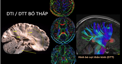
MRI khuếch tán theo hướng và bó sợi thần kinh (DTI/DTT)
5. Magnetic resonance imaging procedure of nerve fiber bundles
5.1 Preparation before the procedure To perform magnetic resonance imaging of nerve fiber bundles to diagnose invasive brain tumor pathology, it is necessary to prepare:Implementation team:
Electroradiologist. Optical technician. Nursing. Means of use:
Resonant angiogram from 1.5 Tesla or more. Film printer. Movie. Image storage system. Medicines: prepare sedatives.
Common medical supplies:
10ml syringe. Cotton, gauze, gloves, sterile bandages. Distilled water (physiological saline). Medicine boxes, first aid tools to prevent contrast drug accidents. Patients need to prepare:
No need to fast. Prepare a copy of the clinician's request for a scan, with a clear diagnosis or complete medical record (if necessary). The procedure is explained in advance to work well with the doctor. Check for contraindications. Change the clothes of the MRI room according to the instructions, remove the contraindicated items.
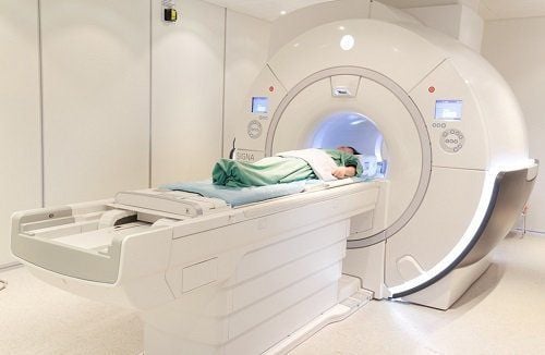
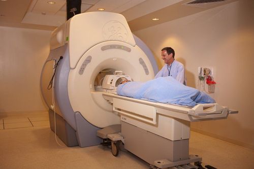
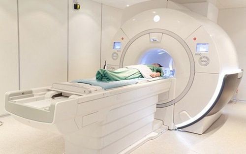
Hệ thống chụp mạch cộng hưởng từ 1,5 Tesla trở lên cần có trong chụp cộng hưởng từ các bó sợi thần kinh
Patient position
Patient is instructed to lie on their back on the shooting table. Select and position the receiver coil. Move the table into the machine bay and position the shooting area. Technique
Locate the area to capture. Select pulse sequences suitable for diagnostic purposes, typically T1, T2 and FLAIR. Customizable cutting directions include: landscape (horizontal), horizontal (horizontal) and vertical (vertical). Perform the usual diffusion pulse sequence, calculate the apparent diffusion coefficient map (ADC map) to assess the extent of the lesion. Note: Other special pulse sequences may be performed for a specific diagnosis of the brain condition (if needed). For example, the T2* pulse sequence helps to identify bleeding in the lesion, the Diffusion pulse helps to assess the stage of infarction, the necrotic substance in the lesion, and differentiates necrotic tumor or brain abscess.
Select the appropriate pulse sequence to capture the nerve fiber bundle (tensor), choose the direction depending on the direction of the axon to be examined. Run each pulse in turn and process the resulting images on the workstation screen, selecting images with clear medical conditions for film printing. Images need to ensure clarity, no vibration, blur and full pulse sequences, maximum support for analysis and diagnosis. Based on the resulting images, the doctor analyzes the results and makes a diagnosis. Note: During magnetic resonance imaging of nerve fiber bundles to diagnose invasive brain tumors, it is necessary to monitor the patient's pulse, blood pressure, and consciousness through the camera and electrocardiogram on the screen.
After the procedure is over, let the patient rest for 30 minutes in the waiting room for further monitoring.
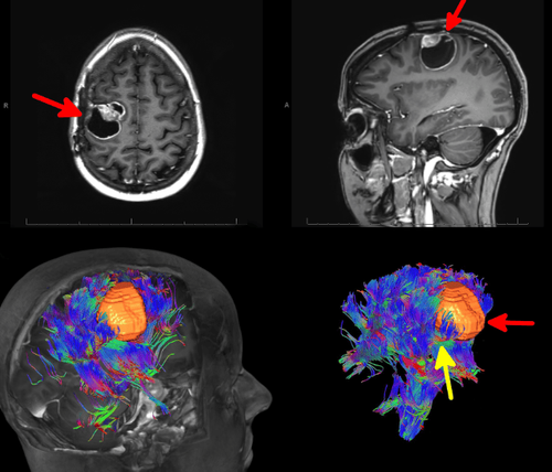
Hình ảnh thu được sau khi chụp cộng hưởng từ các bó sợi thần kinh
Doctor Hai has more than 20 years of experience in the field of diagnostic imaging, especially in the field of multi-segment computed tomography, magnetic resonance. Currently, the doctor is working at the Department of Diagnostic Imaging and Nuclear Medicine - Vinmec Times City International General Hospital.
To register for an examination at Vinmec International General Hospital, you can contact the nationwide Vinmec Health System Hotline, or register online HERE.


