This is an automatically translated article.
Posted by Doctor Vu Thi Hanh - Radiologist - Radiology Department - Vinmec Hai Phong International General Hospital
Magnetic resonance imaging (MRI) is not only the most modern, fast and accurate diagnostic method today, but this imaging method also plays a very important role in the examination and treatment of diseases. related to the prostate gland in men.
1. What is Prostate Magnetic Resonance?
Prostate magnetic resonance is a non-invasive technique used for imaging in the examination and treatment of diseases. Accordingly, magnetic resonance uses strong magnetic fields, RF waves and computers to create detailed images of structures inside the body. However, magnetic resonance does not use X-ray radiation.
Detailed magnetic resonance imaging allows doctors to examine and detect disease, the images can be reviewed on a computer screen, sent by email. copy, print or copy to CD or upload to servers.
Multi-parametric MRI is an advanced form of imaging. It uses three magnetic resonance techniques to provide anatomical images and information about prostate function.
Multi-parametric MRI evaluates water molecule movement (called water diffusion) and blood flow (called perfusion imaging) in the prostate gland. This helps the doctor tell the difference between diseased and normal prostate tissue.

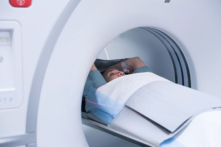
Chụp MRI giúp chẩn đoán bệnh liên quan đến tiền liệt tuyến
2. Purpose of Prostate Magnetic Resonance Imaging
Magnetic resonance to evaluate prostate cancer and see if it is confined to the prostate. Mp-MRI provides information on water molecule distribution and diffusion and blood flow through the prostate. This helps determine if cancer is present and, if so, if it is invasive and if it has spread.
In addition, magnetic resonance of the prostate is used to evaluate for other prostate problems, including:
Infection (prostatitis) or prostate abscess. Benign prostatic hypertrophy Complications after pelvic surgery.
3. Preparing for Prostate Magnetic Resonance Imaging?
Prostate magnetic resonance examination can use abdominal coil or rectal coil, the rectal coil is very close to the prostate helping to create more detailed images, it also allows your radiologist to diagnose your image more accurate injury assessment. In addition, the doctor can perform combined techniques such as magnetic resonance spectroscopy (MRC), diffuse magnetic resonance imaging (DWI), contrast-enhanced magnetic resonance imaging, to provide more information about the chemical structure. learn. Diffusion of water molecules and perfusion in prostate tissue to help differentiate between diseased and normal prostate tissue.
You may be instructed to change into a hospital gown. Or you may be allowed to wear your own clothing if it's not metal.
Before the contrast injection, you may be asked if you have asthma or are allergic to the iodine contrast agent, medication, food, or environment. Magnetic resonance exams often use a contrast material called gadolinium. Gadolinium may be used in patients with an iodine contrast allergy. A patient is less likely to be allergic to gadolinium contrast than to iodine contrast. However, even if the patient has a known allergy to gadolinium, it can be used after appropriate prophylactic administration.
You give the technician or radiologist if you have any serious health problems or have recently had surgery. Severe kidney disease, for example, may require the use of specific gadolinium contrast agents that are considered safe for patients with kidney disease. You may need blood tests to determine if your kidneys are working properly.
If your magnetic resonance examination uses a rectal coil, let the technician know if you are allergic to latex. If so, the technician will cover the endometrial coil with a latex-free condom. To prepare for an MRI with an endometrial coil, eat light meals the day before and the day of your test. This will help make scroll insertion easier. Your doctor may ask you to use an enema before the exam to help clear out your bowels.
If you have claustrophobia (fear of closed spaces) or anxiety, you may want to ask your doctor for a mild sedative before your exam.
Leave all jewelry and other accessories at home or remove them before the MRI. Metallic and electronic devices can interfere with the magnetic field of the MRI room and they should not be brought into the imaging room. These items include:
Jewelry, watches, credit cards and hearing aids, all of which can be damaged. Pins, hairpins, metal zippers, and similar metal items, can distort the MRI images. Dental screws. Pens, pocket knives and eyeglasses. Body piercing. Cell phones, electronic watches and tracking devices. In most cases, magnetic resonance testing is safe for patients with metal implants, with some exceptions. People with the following implants may not be scanned and should not enter the MRI scanning area without first having a safety assessment:
Some cochlear implants Certain types of clips are used for aneurysms brain Certain types of metal coils placed in blood vessels Some older defibrillators and pacemakers Tell the technician if you have medical or electronic devices in your body. These devices may interfere or pose a risk during the examination. Many implantable devices will have a book that explains the MRI risks for that particular device. If you have the manual, give it to your healthcare provider before planning your visit.
Metal objects used in orthopedic surgery generally pose no risk in MRI. However, a recently placed artificial joint may require the use of a different imaging exam.
Tell the technician or radiologist about any shrapnel, bullets, or other metal that may be in your body. Foreign objects near and especially in the eye are important because they can move or heat up during the scan and cause blindness. The dye used in tattoos may contain iron and may heat up during an MRI scan, which is rare. Fillings, braces, eyeshadows and other cosmetics are generally unaffected by magnetic fields. However, they can distort images of the face or brain area.

Các thiết bị điện tử đều không được đem vào khi chụp MRI
4. How to perform Magnetic Resonance Imaging of the Prostate
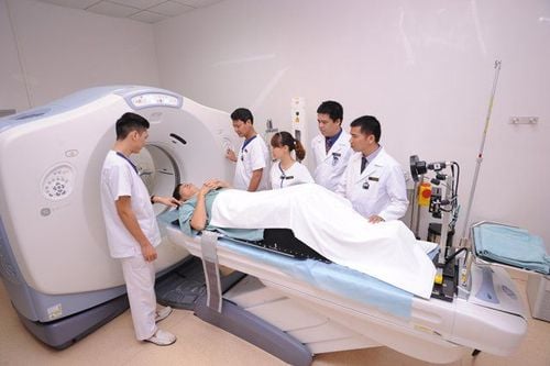
MRI examination can be done on an outpatient basis. The patient will be positioned on a movable examination table. Straps and washers can be used to keep them in place while shooting.
Capture coils capable of sending and receiving radio waves can be placed around or next to the area of the body being scanned. An MRI exam usually involves several runs (sequences), some of which can last several minutes.
Examination of the patient can use coils placed in the rectum. If so, a nurse will place a disposable cover over the coil. The nurse will lubricate the assembly and insert the coil a short distance into the rectum. Once inserted, the nurse inflates the round balloon that sits around the coil and holds it in place throughout the exam. When the test is done, the doctor deflates and removes the coil.
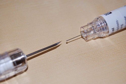
If contrast is used, the doctor, nurse, or technician will place an intravenous line in a vein in your hand or arm that will be used to inject the contrast material. The patient will be placed on the magnet of the MRI machine. The technician will perform the capture technique from a computer in the control room.
If contrast is used during the exam, it will be given into an intravenous (IV) line after the first series of scans. Multiple images will be taken during or after the injection.
Once the examination is complete, the patient may be asked to wait while the radiologist examines the images if more necessary. Accordingly, the entire examination usually takes 45 minutes or less.
Pulmonary resonance imaging may be performed during a prostate exam. This can add about 15 minutes to the total visit time.
5. Benefits versus risks of prostate magnetic resonance imaging
5.1 Benefits of prostate magnetic resonance imaging:
MRI is a non-invasive imaging technique that does not involve radiation exposure. MRI images of soft tissue structures of the body are clearer and more detailed than with other imaging methods. This detail makes MRI a valuable tool in the diagnosis and early assessment of the extent of tumors, such as prostate cancer. MRI has been shown to be valuable in the diagnosis of a wide range of conditions, including cancer. It is also useful in the diagnosis of conditions such as benign prostatic hypertrophy, inflammation, and prostate abscess. Multiparameter magnetic resonance imaging (Mp-MRI) helps differentiate between low-risk and high-risk prostate cancer. It also helps identify cancer that has spread beyond the prostate gland. MRI can detect abnormalities that may be obscured by bone using other imaging methods. The gadolinium MRI contrast material is less likely to cause allergic reactions than the iodine-based contrast materials used for X-rays and CT scans.
5.2 Risks of prostate magnetic resonance imaging:
An MRI exam poses almost no risk to the patient when safety guidelines are followed. High doses of sedation can be dangerous. However, your vital signs will be monitored with a monitor to reduce this risk. Strong magnetic field is not harmful. However, it can cause implanted medical devices to malfunction or cause image distortion. Nephrogenic systemic fibrosis is a recognized, but rare, complication associated with the administration of gadolinium contrast media. It usually occurs in patients with severe kidney disease. Your doctor will carefully evaluate your kidney function before considering contrast injection. There is a very slight risk of allergic reactions if contrast materials are used. Such reactions are usually mild and controlled with medication. If you have an allergic reaction, your doctor will be available to assist immediately.

Chụp MRI rất an toàn, hiếm khi gặp rủi ro
6. What are the limitations of MRI of the prostate?
In addition to the benefits, prostate MRI also has the following limitations:
High quality images depend on the patient's ability to remain still and follow instructions to hold their breath during the scan. Implants and other metal objects can make it difficult to get a clear picture. Patient movement can have a similar effect. To avoid confusing any bleeding with cancer, your doctor may wait six to eight weeks after a prostate biopsy to perform a prostate MRI. MRI is generally more expensive and may take longer to perform than other imaging methods. Vinmec International General Hospital has applied magnetic resonance technology to medical examination and treatment, and image diagnosis. In particular, the prostate magnetic resonance imaging technique at Vinmec is performed methodically and according to standard procedures by a team of highly skilled doctors and nurses, modern machinery system, thus giving accurate results. , contributes significantly to the identification of disease and disease stage, diagnosis and screening of prostate cancer. From there, come up with the most effective treatment plan for the patient.
If you have a need for medical examination by modern and highly effective methods at Vinmec, please register here.


