This is an automatically translated article.
The article was written by Specialist I Tran Cong Trinh - Radiologist, Department of Diagnostic Imaging - Vinmec Central Park International General Hospital.Magnetic resonance imaging of the abdominal aorta has a fairly wide range of uses, fulfilling its role in the diagnosis and treatment of many different pathologies of the abdominal aorta.
1. What is Magnetic Resonance Aorta (MRA)?
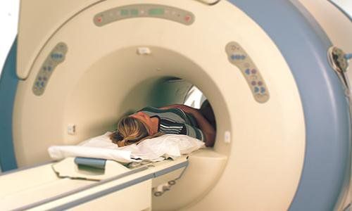
Magnetic resonance imaging (MRI) is a process that uses a strong magnetic field, electromagnetic waves and computer processing systems to create detailed images of body parts, soft tissues, bones. , and other visceral structures.
Magnetic resonance angiography (MRA) of the abdominal aorta is a magnetic resonance imaging procedure that focuses on the abdominal aorta, this procedure may or may not use contrast agents depending on the individual patient. , to produce detailed images of the abdominal aorta, thereby helping to detect the pathology of the abdominal aorta.
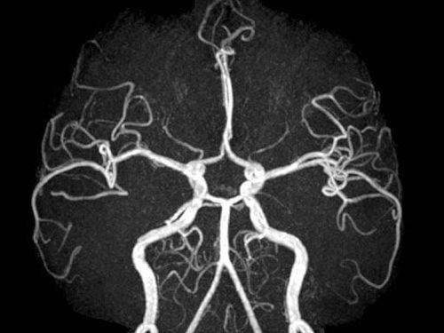
Hình ảnh chụp động mạch chủ bụng có thuốc tương phản
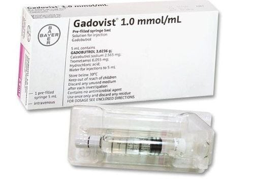
In some cases, the doctor will order more contrast dye. Contrast used in magnetic resonance imaging is less likely to cause allergic reactions than contrast material used in CT scans. Previously, patients could have CT Scan to diagnose abdominal aortic diseases at a lower cost.
However, abdominal aortic magnetic resonance imaging has advantages such as: the patient is not exposed to X-rays as when computed tomography, which helps to reduce the risk of unwanted effects.
2. Indications for abdominal aorta magnetic resonance imaging for which pathology?
Indications for abdominal aortic magnetic resonance imaging include:
Detection of abnormalities such as aneurysms, arterial dissection in the abdominal aorta, or in the branches of the abdominal aorta. Detection of atherosclerotic disease in the abdominal aorta. Assess the degree of narrowing, size, and location of blood vessels involved in vascular disease, supporting the treatment plan of vascular intervention.

Chụp cộng hưởng từ động mạch chủ bụng giúp kiểm tra và phát hiện bất thường mạch máu
Diagnosis of congenital vascular abnormalities in children. An alternative to computed tomography of the abdominal aorta when iodinated contrast agents are contraindicated.
3. Contraindications to abdominal aorta magnetic resonance imaging
In general, MRI of the abdominal aorta is a safe, non-invasive, and radiation-free technique. However, there are some cases where MRI should not be taken, such as:
People with claustrophobia because during the scan, the patient is asked to lie still in the machine's compartment for 45 to 60 minutes. People with claustrophobia may find it difficult to cooperate. The patient harbors foreign bodies inside the body, including: pacemakers, cochlear implants, and certain types of clips used to treat brain aneurysms. Magnetic fields can affect the operation of these devices and conversely they can be the cause of image quality degradation. New-generation artificial substitutes are often provided by the manufacturer with a description of the risks associated with MRI in their manuals. Patients need to discuss carefully with their doctor before deciding to perform.
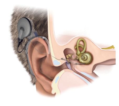
Chống chỉ định chụp MRI với bệnh nhân cấy ốc tai
4. Procedure for performing abdominal aortic magnetic resonance imaging
MRI of the abdominal aorta should be performed according to the correct procedure to ensure the safety of the patient as well as the effectiveness of the device, including the following steps:
Counseling and explaining to the patient how to performed, the risk of abdominal aorta magnetic resonance imaging. Instruct the patient not to fast before the scan. Extract information from patients to rule out contraindications. Patients and relatives entering the MRI room are asked to remove jewelry before entering. During the imaging process, the patient needs to follow the technician's instructions such as keeping the posture, inhale, exhale, .. When the machine is operating, it will make a lot of noise, so the patient is usually equipped with noise-cancelling equipment such as earpads or noise-cancelling headphones Total shooting time may take 45 to 60 minutes. Vinmec International General Hospital with a system of modern facilities, medical equipment and a team of experts and doctors with many years of experience in examination and treatment, patients can rest assured that they will be examined and treated with confidence. treatment at the Hospital.
To register for examination and treatment at Vinmec International General Hospital, you can contact Vinmec Health System nationwide, or register online HERE.


