This is an automatically translated article.
The article was consulted with Dr., Dr. Tran Nhu Tu - Dean and Master, Doctor Nguyen Van Huong - Department of Diagnostic Imaging - Vinmec Danang International HospitalPelvic organ prolapse occurs when the muscles and ligaments that support the pelvic organs are weakened, this condition will get worse over time, if not diagnosed and treated early. Magnetic resonance imaging can assess the prolapse of the pelvic organs.
1. Pelvic Magnetic Resonance Imaging
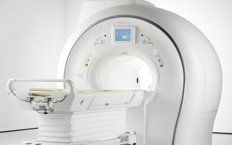
Magnetic resonance imaging, also known as MRI, is a modern medical imaging technique that uses radio waves and magnetic fields, not X-rays. Magnetic resonance imaging has high contrast. , good anatomical detail allows accurate detection of lesions morphological, and structural parts in the body.
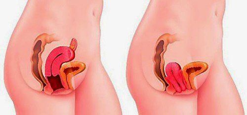
Pelvic organ prolapse is the retraction of one or more pelvic organs from their normal anatomical position, due to damage and weakening of the fascia and ligamentous structures that support the pelvic floor. The disease is common in women, especially in women with multiple births. Although not life-threatening, it greatly affects daily activities. Therefore, early diagnosis and intervention is necessary to improve the patient's quality of life.
Pelvic floor dynamic magnetic resonance imaging is one of the diagnostic measures that allows to better assess the condition of the pelvic floor, early diagnosis of genital prolapse, disorders of fecal outlet obstruction... pelvic organs.
2. Indications and contraindications
2.1 Indications Evaluation of pelvic organ prolapse in patients with symptoms of genital prolapse, urinary incontinence. Find the cause of non-tumor fecal obstruction. 2.2 Contraindications Absolute contraindications
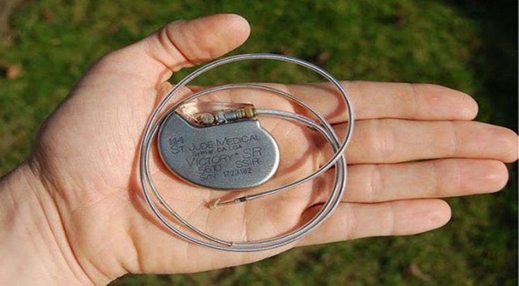
Patients wear electronic devices such as: pacemakers, defibrillators, cochlear implants, automatic subcutaneous drug injection devices, Neurostimulator... Clips metal vascular surgery, intracranial, orbital, less than 6 months. Severe patients need resuscitation equipment next to the body Relative contraindications
Metal surgical forceps > 6 months Patient is afraid of the dark or alone
3. Steps to take
3.1 Preparation Some facilities need to be prepared for magnetic resonance imaging to evaluate pelvic organ prolapse such as:
Resonance angiogram machine from 1 Tesla or more Image storage system, film and printer movie Some drugs are used such as: tranquilizers, contrast agents and antiseptics for skin, mucous membranes, gel (ultrasound). Patients do not need to fast. Before performing, the patient should be thoroughly explained about the procedure to coordinate with medical staff. Instruct the patient to change into the clothes of the MRI room and remove contraindicated items on the body.
In addition, the patient needs an enema to clean the rectum.
3.2 Implementation Steps:
Prepare, position the patient
Inject the gel and position the patient on the patient's side (T) and the ultrasound gel is injected into the rectum (+/- vagina) ) until there is a feeling of defecation (about 200-500mL of ultrasound gel). Have the patient lie down on the scanner in the Fowler position, gently flex the knee with the sandbag under it, and place the receiver coil on the pelvis Protect the patient's ear by covering and warming Selecting and positioning the receiver Di Move the table into the magnetic field of the machine and locate the shooting area Technique
Positioning Capture The machine automatically scans and displays 03 positioning images on 3 planes (cross section, vertical vertical and horizontal plane) Survey the back area o-rectal in longitudinal plane with longitudinal T2-weighted pulses based on transverse and frontal plane localization. Examine the pelvis in the frontal plane with a horizontal HASTE T2 pulse along the anal canal axis on a vertical T2-weighted image. Take 01 vertical image right in the middle of T2 HASTE and stand along 1 slice as a standard for recording the umbilicus - anal push. Based on the transverse image, the slice is right in the middle of the anal canal and the cross-sectional image is parallel to the vertical plane of the body. Have the patient perform anal retraction (pinch) and record by pulse Cine Sagittal Trufisp on the vertical image in the middle 1 second / 1 image for about 15 seconds. For the patient to continue to perform defecation manipulation, ultrasound gel and Cine Sagittal Trufisp recording similar to when pinching the anus for about 45-60 seconds Cross-sectional examination of the pubic-rectal muscle on transverse pulse T2 HASTE in neutral position. For the patient to do the Valsalva maneuver, examine the transverse image of the pubic rectum on the T2 transverse pulse HASTE VALSALVA. horizontal Trufisp in about 30 seconds (1 image / 1 second) Technician prints film, transfers images to doctor's workstation Doctor analyzes images, ejection parameters and diagnoses Results judgment
Obtain Receive images of anatomical structures of the anorectal region. Assess the kinematics of the pelvic floor, anatomical structures of the anorectal region and the kinetics of rectal expulsion. Magnetic resonance imaging to evaluate prolapse of the pelvic organs is a diagnostic method to help detect abnormalities in the position of the organs compared to the original anatomical structure. Early diagnosis helps patients receive timely intervention, improving quality of life.
Vinmec International General Hospital is one of the hospitals that not only ensures professional quality with a team of leading medical doctors, modern equipment and technology, but also stands out for its examination and consultation services. comprehensive and professional medical consultation and treatment; civilized, polite, safe and sterile medical examination and treatment space.
Please dial HOTLINE for more information or register for an appointment HERE. Download MyVinmec app to make appointments faster and to manage your bookings easily.


