This is an automatically translated article.
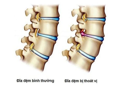
The article is professionally consulted by Dr. Pham Quoc Thanh - Radiologist - Radiology Department - Vinmec Hai Phong International General HospitalSpinal disc herniation has a significant impact on the quality of life of patients. Magnetic resonance imaging to diagnose spinal disc herniation is very important, helping patients to be detected early and intervene promptly.
1. Overview of spinal hernia pathology
The spine is composed of vertebrae and discs stacked on top of each other, there are about 32 - 34 vertebrae, divided into 5 segments arranged from top to bottom. The spine performs the following tasks: protecting the spinal cord, moving, bearing the body's load and protecting internal organs.
Lumbar spine pain is caused by many causes, in which typical can be mentioned:
Acute herniation or disc protrusion due to incorrect posture during work; Soft tissue damage from overwork; Joints and discs degenerate, causing herniated disc disease and spinal instability. Depending on the duration of pain, back pain is divided into: acute and chronic. Cases of low back pain less than 4 weeks are considered acute, cases of pain that appear slowly and gradually increase over time over 4 weeks are called chronic.
2. Diagnostic methods for spinal hernia
2.1 Conventional X-ray Through some pictures of conventional X-rays such as: scoliosis, loss of spinal curvature, intervertebral stenosis... can locate the hernia. In addition, conventional X-ray also helps to identify other injuries of the spine such as waist defect, spinal instability, spondylolisthesis...
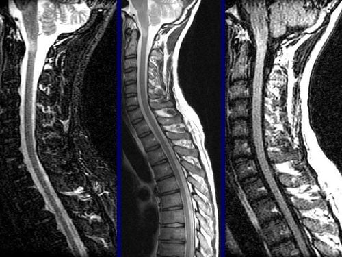
2.2 Magnetic resonance imaging (MRI) Magnetic resonance imaging allows to identify location, hernia morphology, number of hernia floors. This is considered the most modern and accurate imaging method in the diagnosis of lumbar disc herniation.
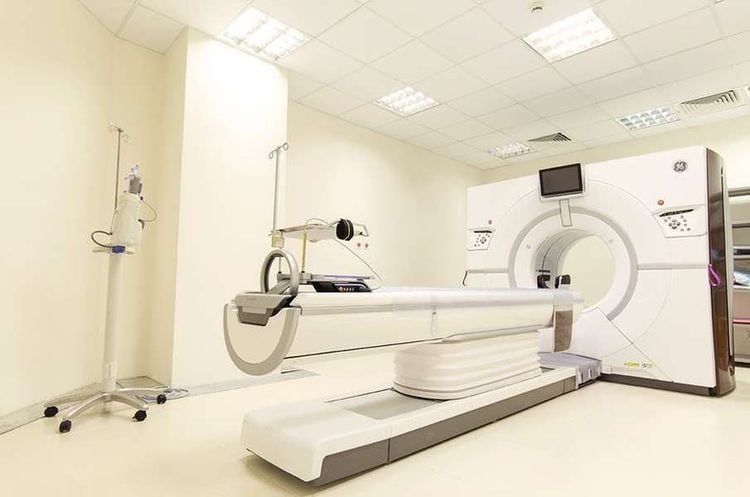
2.3 Computed tomography combined with contrast root encapsulation In case the patient is suspected of disc herniation but unable to undergo magnetic resonance imaging, computed tomography combined with contrast root capsule will be indicated. This technique allows to determine the location and extent of the hernia accurately with high sensitivity.
3. When is magnetic resonance imaging of the spine needed?
It is necessary to visit a medical facility as soon as the following symptoms appear:
After carrying heavy objects, you have back pain or back pain for a long time with no known cause; Loss of bladder or bowel control; Have a history of cancer; Numb limbs; Frequent pain spreading from the back to the buttocks, thighs and legs, affecting daily life.
4. When is a lumbar spine scan indicated?
Diagnostic imaging by magnetic resonance imaging of the lumbar spine will be indicated in the following cases:
Evaluation of anatomical abnormalities of congenital pathologies related to the lumbar and spine; Diagnosis of diseases in the spine and low back such as: Herniated or degenerative discs; assessment of nerve roots, spinal cord compression; Diagnosis of spinal tumor, early-stage spinal metastases, surrounding nerves and soft tissues; Detect birth defects of the spine; Evaluation after spinal cord injury to detect damage and abnormalities in discs, bones, ligaments, spinal cord; Evaluation of a compressed, inflamed nerve; Detecting tuberculosis of the spine; Diagnosis of spinal diseases such as myelitis, myeloma, white matter; Diagnosis of diseases in the spinal canal such as hematomas, tumors in the spinal canal.
5. Benefits of lumbar spine magnetic resonance imaging
Like X-ray, magnetic resonance imaging of the lumbar spine has many outstanding advantages in imaging diagnosis. However, the image quality obtained from magnetic resonance imaging is many times clearer. People who have had back pain for a long time but X-ray or CT scan is not clear, and it is difficult to identify the pathology, they should have an MRI. Because of that, this method is not only applied to bone and joint imaging but also applies to many different parts of the body.
Besides, magnetic resonance also helps diagnose lesions in the vertebral body, spinal cord, disc and soft tissue around the spine. The spine images obtained from MRI are clear and detailed.
6. Notes when taking magnetic resonance imaging of the spine
When taking magnetic resonance imaging of the spine, it is important to note:
Do not carry mobile phones and metal objects in your body during MRI scans of the lumbar spine because the magnetic waves of the scanner will affect these devices, causing harm to people. sick; Cases requiring anesthesia for MRI should fast for 5-6 hours before the scan. The rest of the cases do not need to do this; For patients who require an artificial pacemaker, defibrillator, pacemaker or hearing aid, or a metal diabetic subcutaneous auto-injector, no spinal resonance imaging should be performed; Patients only take MRI of the spine when prescribed by a specialist so that they can monitor their health and receive timely treatment when necessary. In order to diagnose spinal hernia in a short time, have correct results and support effective treatment, patients should perform this technique at reputable medical facilities with modern MRI machines and systems. Standard studio. The team of doctors performing the technique needs to be well-trained, highly qualified and experienced to be able to read the film accurately, thereby helping the patient get the best treatment regimen.
Please dial HOTLINE for more information or register for an appointment HERE. Download MyVinmec app to make appointments faster and to manage your bookings easily.


