This is an automatically translated article.
The article is expertly consulted by Master, Doctor Nguyen Thi Mai Anh - Doctor of Radiology - Department of Diagnostic Imaging and Nuclear Medicine - Vinmec Times City International General Hospital. Doctor Nguyen Thi Mai Anh has nearly 10 years of experience in the field of diagnostic imaging, especially in imaging breast and thyroid cancer.Ultrasound is an imaging method widely indicated in the diagnosis of many liver-related diseases, the most prominent of which is cirrhosis. So when is the liver ultrasound indicated and what are the liver cirrhosis images on ultrasound?
1. What is cirrhosis?
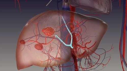
Cirrhosis is a consequence of untreated liver damage for a long time, liver cells gradually become fibrosis over time, causing liver function to be reduced or completely lost.These fibrous cells will displace and gradually replace normal liver cells and thus the liver will also gradually lose its main functions. Common causes that can cause cirrhosis include: hepatitis caused by alcohol abuse, hepatitis caused by hepatitis B virus, hepatitis C virus or drug and chemical poisoning...
2. Dangerous complications of cirrhosis
Cirrhosis usually goes through 4 stages, if in the early stages, cirrhosis often does not cause obvious symptoms, but this is the time of effective treatment, prognosis for complete recovery and no sequelae.However, because there are no specific signs, most patients with cirrhosis are diagnosed incidentally (when doing a routine physical examination or when treating another disease) or when the disease is already at an advanced stage. decompensation and severe complications.
At that time, the treatment ability was very low, the liver cirrhosis lost function and the patient died very quickly because of the following dangerous complications:
Gastrointestinal bleeding: Cirrhotic tissues increase venous pressure portal, causing the portal-host junction system to increase pressure and relax, to a certain limit it will burst, causing upper gastrointestinal bleeding due to rupture of esophageal varices, which can be life-threatening due to excessive blood loss. a lot of. Edema, ascites: Increased portal pressure accompanied by hypoproteinemia due to decreased production causes edema (in the lower extremities) and ascites (extra-abdominal fluid). Cirrhotic encephalopathy: The liver plays a role in both production and elimination of toxins. Therefore, cirrhosis causes the liver to gradually lose its functions, unable to eliminate toxins from the body, causing the body to accumulate high concentrations of toxins, these substances go to the brain, causing encephalopathy syndrome, coma. liver and rapidly died. Kidney failure Infection.
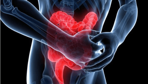
Các mô xơ gan làm tăng áp lực tĩnh mạch cửa, làm hệ nối cửa - chủ tăng áp lực theo và giãn ra, đến một giới hạn nhất định sẽ vỡ, gây xuất huyết tiêu hóa trên
3. What is liver ultrasound?
Liver ultrasound is a method of using ultrasound waves through a special transducer placed on the patient's abdomen to observe images of the liver.Liver ultrasound is a modern method, easy to perform and safe for patients because it is non-invasive. High reliability and accuracy of ultrasound, can be applied to all objects.
However, liver ultrasound requires the person performing the ultrasound to have high expertise and experience to record liver abnormalities.
4. When is liver ultrasound indicated?
When the patient has abnormal symptoms and signs that suspect liver problems, specifically cirrhosis, the doctor will assign the patient to an ultrasound of the liver. Common signs and symptoms in cirrhosis patients depending on the stage include:Jaundice, yellow eyes; Patients feel tired, sluggish, lose weight quickly; Anorexia, anorexia, loss of appetite; Digestive disorders ; Blood clotting disorders: Easy bruising and bleeding; Leg swelling or ascites (bulging of the abdomen due to body fluids).
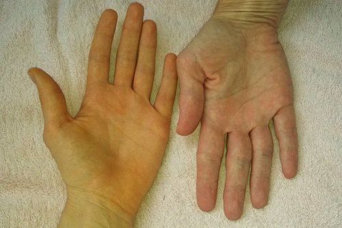
Vàng da, vàng mắt là triệu chứng dễ nhận biết ở bệnh nhân xơ gan
5. What is cirrhosis of the liver on ultrasound?
Usually, some images of cirrhosis on ultrasound include:Concave liver margin, rough liver parenchyma, appearance of small nodules on the liver surface; Combined atrophy and hypertrophy of the lobes of the liver: the outer lobe, caudal lobe increase in size and the right lobe (especially the posterior segment) decreases in size. Record abdominal fluid;
6. Ultrasound technique for measuring liver elasticity
In the process of monitoring, treatment and prognosis of cirrhosis, it is extremely important to diagnose the degree of fibrosis of liver cells. For the most accurate assessment, a liver biopsy is required, but this technique is invasive and carries many risks and complications for the patient.Therefore, to meet the above requirements, the liver ultrasound technique to measure liver tissue elasticity was developed and gradually became the first priority in determining the degree of liver fibrosis and accompanying liver tumors. if any.
This liver ultrasound technique is also known as Fibroscan with the advantage of being non-invasive, easy to perform, repeatable many times and giving high accuracy results. Currently, liver ultrasound measuring liver tissue elasticity is used more and more widely in the diagnosis, monitoring and prognosis of cirrhosis.
7. What to prepare when performing liver ultrasound?
Liver ultrasound for diagnosis of cirrhosis is a simple, easy-to-implement imaging method and does not require any special preparation prior to ultrasound.Ideally, the patient should fast before the liver ultrasound for about 8-12 hours to remove the interference images on the gastrointestinal tract that affect the results of the liver ultrasound.
When conducting an ultrasound of the liver, the doctor will apply a special gel on the abdomen where the liver is located, then use the probe to observe the entire liver through the black and white image displayed on the screen. Based on this image, the doctor will make a conclusion about the patient's liver condition.

Điều kiện lý tưởng nhất là bệnh nhân nên nhịn ăn trước siêu âm gan khoảng 8 – 12 giờ
Please dial HOTLINE for more information or register for an appointment HERE. Download MyVinmec app to make appointments faster and to manage your bookings easily.