This is an automatically translated article.
The article was written by Specialist Doctor II Khong Tien Dat - Radiologist, Department of Diagnostic Imaging - Vinmec Ha Long International General Hospital.The technique of computed tomography of the spine aims to create images of the lumbar spine with a computed tomography machine to evaluate lesions of the bones, discs, spinal canal, spinal cord and adjacent components..
.
.
1. Indications and contraindications
Indications
Traumatic pathology, tumors, inflammation of the bones and soft parts of the lumbar spine.
Contraindications
No absolute contraindications Relative contraindications for pregnant women

Phụ nữ mang thai không nên chụp cắt lớp vi tính cột sống thắt lưng
2. Prepare to shoot
+ Performer:
Specialist doctor, Electro-optical technician, Nursing equipment, CT scanner Film, sand, image storage system + Patient:
The patient is thoroughly explained about the procedure to coordinate with the physician. Remove earrings, necklaces, and hairpins, if any. In case the patient is too excited, cannot lie still, sedation is required. There is an order form for computed tomography from the examining clinician.
3. Steps to conduct computed tomography
Patient's position: Place the patient in the machine frame, lie on his back, with the shoulders as low as possible, with both hands raised up along the body axis. Hold your breath and do not swallow during the examination. Technical implementation:
The spine in the examination area is localized in the vertical direction. Get the positioning shape in the vertical orientation. Set the imaging program according to clinical requirements. Directional slices of the discs can be used to evaluate for herniated disc pathology. After taking the photo, use software that allows to process the image after taking it to reveal the lesion.
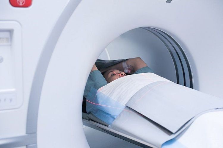
Bệnh nhân nằm trong khung máy chụp cắt lớp vi tính
4. Evaluation of results
Evaluation of vertebral body injuries such as: vertebral body rupture, vertebral collapse, vertebral body slip, especially the image of displaced wall lesions behind the vertebral body (because of the very high risk of compression of the vertebral body and the tundra), the posterior arch damage, traumatic hematoma and especially signs of disc herniation, soft tissue lesions of the vertebral canal, location of iodine-contrast foreign bodies. Injuries in degenerative spondylolisthesis such as: lateral joint block degeneration, ligament degeneration, degenerative spondylolisthesis, spinal stenosis. Evaluation of congenital abnormalities of the spine.
5. Complications and treatment
No technical accidents. Some errors may require re-implementation of the technique such as: the patient does not remain motionless during the film, does not clearly reveal the image. Vinmec International General Hospital is one of the hospitals that not only ensures professional quality with a team of leading doctors, modern equipment and technology, but also stands out for its examination and consulting services. and comprehensive, professional medical treatment; civilized, polite, safe and sterile medical examination and treatment space.
Customers can directly go to Vinmec Health system nationwide to visit or contact the hotline here for support.


