This is an automatically translated article.
The article is professionally approved by Master, Doctor Nguyen Viet Thu - Doctor of Radiology and Nuclear Medicine - Department of Diagnostic Imaging and Nuclear Medicine - Vinmec Times City International General HospitalToday, respiratory diseases are increasing in both morbidity and mortality (especially lung cancer). Imaging methods are also increasingly diversified, including bronchoscopic ultrasonography, which makes the diagnosis of respiratory diseases easier.
1. Overview of bronchoscopic ultrasound
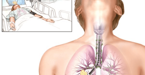
Endobronchial ultrasound (EBUS) is a technique of bronchoscopy using a flexible tube through the mouth or nose, helping doctors observe the respiratory tract to diagnose lung diseases. This procedure allows a biopsy to take samples of tissue, cells, or lung fluid for testing. Ultrasound bronchoscopy is also used in the treatment of certain lung diseases.Bronchoscopy has 2 main types of probes:
Radial probe endobronchial ultrasound (RP-EBUS) for peripheral lung lesions. Convex probe endobronchial ultrasound (RP CP-EBUS) is for mediastinum, hilar nodes or parabronchial tumors. This is considered a routine and first technique to help diagnose lesions in these locations.
2. Indications for ultrasound bronchoscopy
Ultrasonic bronchoscopy will help doctors diagnose respiratory diseases by examining the airways in the lungs or taking a biopsy sample.
Ultrasound bronchoscopy is usually indicated in the following cases:
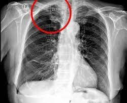
Suspected lung tumor, lymph node, atelectasis. Abnormal findings on X-ray film or other imaging methods. Suspected pulmonary interstitial disease, lung and bronchial infections. The patient has hemoptysis or a cough that persists for more than 3 months of unknown cause. The patient breathes in toxic gases or chemicals. Clear mucus or sputum that is obstructing the airways should be removed. Dilate a blocked or narrow airway. Abscess drainage.
3. Procedure for performing ultrasound bronchoscopy
3.1. Prepare for the procedure
Before performing the procedure, the doctor will explain the procedure and ask the patient to consider and sign the consent form for the procedure.
In some cases blood tests may be needed before the endoscopy. Patients are required to fast for at least 4 hours before the procedure, but can drink water 2 hours before. Patients should not take blood thinners such as Aspirin, Ibuprofen before the procedure. If the patient wears dentures, the denture needs to be removed. The doctor may place a catheter (catheter) into the patient's vein to administer medication to help relax or induce anesthesia if needed. Because the patient may be drowsy after the procedure, it is necessary to have a family member with you or prepare transportation before discharge.
3.2. Ultrasonic bronchoscopy procedure
The bronchoscope with a camera will be inserted into the airways through the mouth or nose into the trachea, leading to the patient's lungs. During the procedure, the majority of patients remain very awake.
Patient is guided into appropriate position. The raised head of the bed helps the patient to have a sitting position, with the head slightly tilted back as if looking up at the ceiling.
Oral endoscopy: The doctor will spray numbing medicine into the patient's mouth and throat to reduce discomfort and nausea when the bronchoscope goes in. Nasal endoscopy: The doctor will place a numbing jelly in one nostril. The patient may have a cough when the bronchoscope begins to enter, but when the anesthetic takes effect, this condition disappears, and the patient will not be able to feel in the bronchoscope. When the patient is fully absorbed, the bronchoscope will be inserted into the lungs.
During the ultrasound bronchoscopy, the patient may need to provide oxygen through a tube inserted into the nose or use an oxygen mask, combined with the use of a heart rate monitor. The doctor can pump saline through the bronchoscope to wash the lungs, biopsy samples of lung fluid, lung cells, and other substances in the alveoli for testing. Your doctor may also place a stent in your airway.
3.3. End of bronchoscopy ultrasound procedure
After the anesthetic wears off, the patient may experience cough reflex, hoarseness or sore throat for 1-2 hours, sore throat for a few days.
Local anesthetic sprayed into the patient's throat can affect the patient's ability to swallow up to 4 hours after the endoscopy procedure. Therefore, the patient must not eat or drink until the swallowing reflex returns.
After bronchoscopy, the patient can be discharged on the same day, depending on the case, the patient can choose to rest overnight in the inpatient treatment area.
Ultrasonic bronchoscopy is currently a highly appreciated technique to help detect respiratory diseases accurately so that there can be timely treatment.
With the desire to bring comfort as well as minimize the impact on health, Vinmec International General Hospital, has applied flexible bronchoscopy technique with modern machinery and equipment, Patients will not feel discomfort during endoscopy. Before the procedure, the patient will be anesthetized by medical staff and carefully controlled the airway to ensure safety and minimize irritation or discomfort for the patient. Next, the doctors will perform endoscopic techniques, check to assess the health status.
The examination process is performed by a team of qualified doctors with many years of experience combined with modern equipment and machines. Therefore, patients can be completely assured of the examination results.
Please dial HOTLINE for more information or register for an appointment HERE. Download MyVinmec app to make appointments faster and to manage your bookings easily.


