This is an automatically translated article.
The article is professionally consulted by Master, Doctor Hoang Thanh Nga - Ophthalmologist - Department of Medical Examination and Internal Medicine - Vinmec Ha Long International General Hospital.
The eye is a visual organ that performs the function of observing, seeing, and receiving images, colors of things transferred to the brain for processing and storage. In particular, the pupil and cornea are one of the important parts to ensure the vision function.
1. Structure of the eye
The eye is a visual organ that performs the function of seeing, observing and retrieving images and colors of things to transfer to the brain for processing and storage.
The outer structure of the eye includes: eyelids, eyebrows, eyelashes, irises and whites. In addition, the pupil, cornea, lens, and retina are the internal components of the eye.
2. Pupil Size
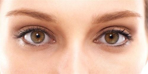
The pupil is the part of the eye, a hole in the center of the iris that allows light to pass through and reach the retina. The pupil is black because light that has passed through has either been directly absorbed by the tissues on the inside of the eye, or absorbed after being diffused by reflection inside the eye and not being able to escape through the narrow pupil.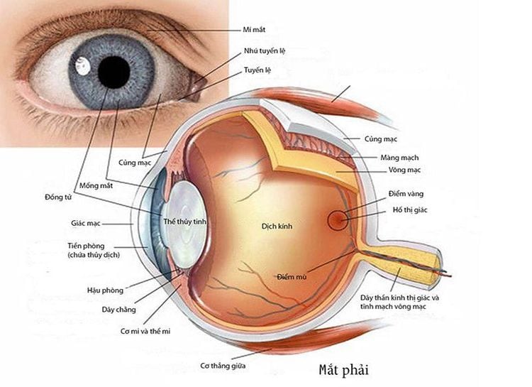
Vị trí của đồng tử trong mắt
The size of the pupil depends on age and light. At the age of 15, the pupil will be at its largest, about 3-8 mm. After the age of 25, the average pupil size will start to decrease, but there is no fixed rate.
Under natural conditions, the pupil will contract gently. When the light is strong, the pupil will constrict to prevent excess light from entering the eye and increase the sharpness of the image. At this point the pupil is about 2-4 mm. The less light reaching the eye, the larger the pupil is and vice versa.
Dilation of the pupil is known as pupillary dilation, usually due to an abnormal cause, or a physiological response of the pupil. It can be caused by injury, substance use, or illness. In contrast, pupillary constriction is the constriction of the pupil. Pupil dilation is used in important brainstem injury testing.

Kích thước đồng tử có thể thay đổi do ánh sáng
3. Corneal Diameter
The cornea is a transparent, non-vascular, very tough, and cone-shaped membrane occupying the anterior 1/5 of the ocular cortex. The diameter of the cornea is about 11mm and the radius of curvature is 7.7mm. The thickness of the cornea is thinner in the central region than in the margin.
The anterior curvature radius of the cornea forms a focusing force of about 48.8D, which accounts for two-thirds of the total refractive power of the eyeball. The cornea has 5 layers from outside to inside: epithelial layer, bowman membrane, parenchyma, Descemet membrane and endothelium.
In short, the cornea and pupil are one of the parts located in the anterior part of the eye, responsible for protecting the eyes against the effects of light, dust,... Elasticity, change in size of the pupil also reflects a non-physiological cause of the body.


