This is an automatically translated article.
Inflammatory myopathies is a less common autoimmune disease than systemic lupus erythematosus, scleroderma, and periarteritis nodule. Typical symptoms of the disease are dermatitis, myositis, and muscle weakness. It usually occurs in middle-aged people between 40 and 60. However, children under 10 years of age can also develop inflammatory myopathy.
1. What is myositis?
Inflammatory myopathies are a group of inflammatory myopathies characterized by muscle weakness. When there is only muscle involvement, it is called polymyositis, when accompanied by damage to the skin, it is called dermatomyositis.
Inflammatory myopathy is a connective tissue disease characterized by muscle inflammation. Some of the causes of the disease can be: infections, drugs, toxins, metabolic disorders, idiopathic inflammatory myopathies, paroxysmal globulinuria syndromes (Consequences of stress in people with metabolic disorders) potential.)
2. Symptoms of dermatomyositis
2.1. Muscle
2.1.1. Physical symptoms
Progressive weakness is considered the most important symptom, mainly in the limbs and shoulder blades. People with myositis may have:
Unable to climb stairs or having difficulty; Inability to raise the seat; Unable to keep the hand steady; Has bilateral symmetry; Trendelenburg gait: Excessive arching of the spine. Muscle pain: 50% of cases have pain, muscle sensitivity.
Slow disease progression.
End stage: Muscle atrophy
The muscles of the head of the face may be atrophied or associated with malignancy. The muscles of the pharynx, larynx, and digestive tract can become atrophied.

Teo cơ là một trong những triệu chứng nguy hiểm của viêm cơ da
2.1.2. Physical symptoms
Detecting the injured muscle position is usually at the base of the limb, showing reduced muscle strength, muscle tension, especially when palpated; Reduced mobility; Signs of "stool" due to weakness of proximal muscles or difficulty standing up while sitting, in severe cases, the patient cannot stand up and walk on his own;
2.2. Skin
2.2.1. Physical symptoms
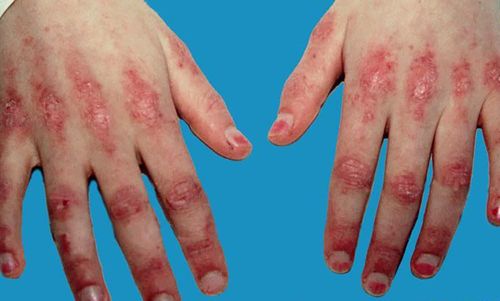
Presence of early skin lesions makes it difficult to diagnose inflammatory myopathy. Maculopapular erythema: Joints, elbows, knees, toes (70%); Appears small maculopapular nodules that gradually enlarge; Color: The rash is red purple, with vasodilatation, scabs. Many maculopapular lesions appear on the hands and feet that can progress to Poikiloderma 60% of inflammatory myopathies present with red face, pale purplish color around the eyelids (Heliotrope), especially in children. Dilated capillaries around the nail area (common in overlap connective syndrome). There are some similar lesions such as Lichen Plan, Duhring, SLE, scleroderma, photodermatitis. Calcinosis: Diffuse calcium deposition under the skin, bones, and muscles, possibly with ulcer complications. Symptoms may appear in the oral mucosa.
2.2.2. Physical symptoms
Typical skin symptoms of inflammatory myopathy are maculopapular erythema (red-purple rash) around the eyes and Gottron's papules in adjacent bony areas. Some other possible manifestations are hair loss, change in nail morphology or subcutaneous calcification.
2.3. Other symptoms
Aching joints (15-30%); Is the esophageal muscle inflamed? Myocarditis (40%); Pulmonary fibrosis (10%); Bowel and stomach cancer; Eyes: Hemorrhage, iridocyclitis, strabismus,...
3. Subclinical index of dermatomyositis
An increase in creatine kinase (CK) levels is an unreliable indicator, although it can be very high. Levels of SGOT, SGPT, LDH, and aldolase may also be elevated. Autoantibodies: Antinuclear antibody (ANA) positive: common in inflammatory myopathies. Anti-Mi-2 antibodies are specific for dermatomyositis but are found in only 25% of inflammatory myopathies. . Anti-Jo-1 antibodies are associated with interstitial lung disease, Raynaud's phenomenon, and arthritis. MRI is not diagnostically helpful, but it can help monitor inflammatory myopathies and guide the best site for muscle biopsies. Electromyography can be helpful, but 15% of cases may still have normal electromyography. Electromyography can also guide the selection of a suitable site for a muscle biopsy. Muscle biopsy: can help diagnose inflammatory myopathy.
4. Treatment of dermatomyositis
4.1. Principles of treatment of dermatomyositis
No specific treatment Need to treat inflammatory myopathy as soon as the disease is diagnosed Principle: anti-inflammatory + symptomatic treatment

Bệnh viêm cơ da cần được chẩn đoán càng sớm càng điều trị hiệu quả
4.2. Commonly used medications
Glucocorticoids: prednisolone, prednisone, methylprednisolone
Usual dose: prednisolone, prednisone or methylprednisolone starting 1-1.5 mg/kg/day orally or by infusion, divided into 2-3 times a day, after 2-4 weeks Administer once a day, maintain treatment for 4-12 weeks and gradually reduce the dose, usually 10-15% of the dose every 2-4 weeks and maintain at a dose of 5-10mg/day or every other day. High dose intravenous (pulse dose): methylprednisolone 500-1000 mg/day, intravenous infusion over 30 minutes, infusion for 3 consecutive days, then return to normal dose High doses are usually indicated for the treatment of acute attacks of cases of severe inflammatory myopathy unresponsive to usual doses. Monitor treatment: blood pressure, bone density, blood sugar, blood calcium levels, blood cortisol, ACTH test, symptoms of peptic ulcer disease. Immunosuppressive drugs: 25% of cases of dermatomyositis combine glucocorticoids with immunosuppressive drugs to control disease symptoms.
Methotrexate: is the drug of first choice Cyclophosphamide: dosage: 0.5-1g IV once a month for 6 months. Monitor treatment: Blood count, platelet count, hematocrit at least once a month during treatment. Mycophenolate mofetil: dose: 2g/day for the first 6 months, 1g/day for the next 6 months, maintenance dose of 0.5g/day for 1-3 years. Monitoring: Liver enzymes 1 month after treatment and every 3 months during the whole treatment. Chloroquine: In cases of skin lesions, chloroquine treatment may be considered. Dosage: 0.25 g/day for 6 months to 1 year. Monitoring: Blood count, eye examination every 6 months during treatment. Rehabilitation and physical therapy: Early initiation improves function and reduces the risk of muscle spasticity. Myositis can affect both adults and children, greatly affecting the health and life of the patient. The disease will be treated faster and more effectively if detected early. To get the earliest examination at Vinmec, you can book an appointment HERE.


