This is an automatically translated article.
The article was professionally consulted with Master, Doctor Nguyen Van Huong - Department of Diagnostic Imaging - Vinmec Danang International General Hospital.Lymphoma is a disease that develops in the cells of the lymphatic system. A blood test or lymph node biopsy is the way to diagnose the disease. To determine if the cancer has spread, a patient may have a bone marrow biopsy, lumbar puncture, and other imaging tests. Treatment depends on the type of lymphoma, the stage of the disease, and the patient's age and overall health.
1. Lymphoma and what you need to know
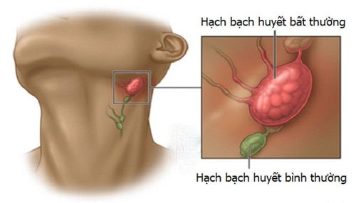
Lymphoma is cancer that develops in the white blood cells (lymphocytes) of the lymphatic system. White blood cells are part of the body's immune system. The lymphatic system consists of a network of small channels similar to blood vessels that circulate fluid (called lymph), the lymph nodes, bone marrow, and several organs in the body are all made up of cells. white blood cells. Lymph nodes have a flat oval shape, they are present all over the body, but they are concentrated in some areas such as the neck, armpits, and groin.There are two main types of lymphoma: Hodgkin (HL) and non-Hodgkin (NHL). Within each category there are several sub-categories. Hodgkin's lymphoma is also known as less common Hodgkin's disease. Non-Hodgkin lymphomas are individual lymphomas that differ in how they work, how they spread, and how they respond to treatment.

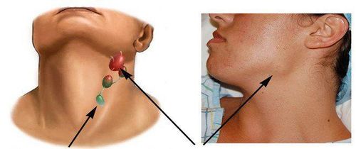
Symptoms of lymphoma may include: swollen lymph nodes in the neck, armpits, groin, unexplained weight loss, fever, night sweats, body itching, fatigue, loss of appetite, cough or difficulty breathing pain in the abdomen, chest or bones, swollen abdomen.
2. Tests, biopsies, and imaging studies in lymphoma assessment
To make a diagnosis, a doctor will begin by taking a patient's medical history and symptoms and performing a physical exam. Your doctor may recommend one or more of the following tests.Blood tests: Usually the white blood cell, platelet, and red blood cell count will be low when the lymphoma has spread to the bone marrow. Blood test results also aid in assessing how well the liver and kidneys are functioning. Lymph node biopsy: A procedure in which part or all of a lymph node is surgically removed for examination under a microscope to look for the presence of lymphoma cells. Other laboratory tests may be performed on the biopsy sample, including molecular genetic testing. Biopsies are often used to determine disease status, disease progression, and determine further tests and treatments. Before the biopsy, the patient needs to do routine tests and imaging tests. The risk of the biopsy procedure is assessed as low risk, the most common risks that can only occur are local bleeding and infection but are very rare. Bone marrow aspiration and biopsy: This is a procedure in which a thin, hollow needle is inserted into the hip bone to remove a small amount of liquid bone marrow and analyze it under a microscope. This procedure is usually done after lymphoma has been diagnosed to determine if the disease has spread to the bone marrow.


Lymphoma is a disease with a good prognosis, if detected early, if not detected and treated promptly, it will lead to severe disease progression and inability to cure
Periodic health check-up is Vinmec's strength Due to good organization, accurate results, continuous monitoring and evaluation of patients, it is possible to detect early systemic abnormalities especially of the lymphatic system. Highly trained and skilled personnel, modern machinery, especially the CT 640 and MRI 3 systems, Tesle scans the whole body for abnormal lymph nodes very well.
Currently, Vinmec International General Hospital has been and continues to be fully equipped with modern diagnostic facilities such as: PET/CT, SPECT/CT, MRI... biology, immunohistochemistry, genetic testing, molecular biology testing, as well as a full range of targeted drugs, the most advanced immunotherapy drugs in cancer treatment. Multimodal cancer treatment from surgery, radiation therapy, chemotherapy, hematopoietic stem cell transplantation, targeted therapy, immunotherapy in cancer treatment, new treatments such as autoimmunotherapy body, heat therapy...
After having an accurate diagnosis of the disease and stage, the patient will be consulted to choose the most appropriate and effective treatment methods. The treatment process is always closely coordinated with many specialties: Diagnostic Imaging, Biochemistry, Immunology, Cardiology, Stem Cell and Gene Technology; Department of Obstetrics and Gynecology, Department of Endocrinology, Department of Rehabilitation, Department of Psychology, Department of Nutrition... to bring the highest efficiency and comfort to the patient. After undergoing the treatment phase, the patient will also be monitored and re-examined to determine whether the cancer treatment is effective or not.
Especially, now to improve service quality, Vinmec also deploys many cancer screening packages that can help customers detect cancer early before there are no symptoms, bringing a better prognosis. treatment and a high chance of recovery.
Please dial HOTLINE for more information or register for an appointment HERE. Download MyVinmec app to make appointments faster and to manage your bookings easily.
Reference source: radiologyinfo.org


