This is an automatically translated article.
The article was professionally consulted by resident Doctor Nguyen Quynh Giang - Department of Diagnostic Imaging and Nuclear Medicine - Vinmec Times City International Hospital.Magnetic contrast-enhanced joint magnetic resonance imaging (MRI) is one of the most common bone and joint imaging tests. Compared with other imaging methods, and thanks to the injection of magnetic contrast agents, MR imaging of joints helps to identify soft and hard tissue structures clearly and accurately, especially in acute conditions. .
1. What is Magnetic Resonance Imaging with Magnetic Contrast?
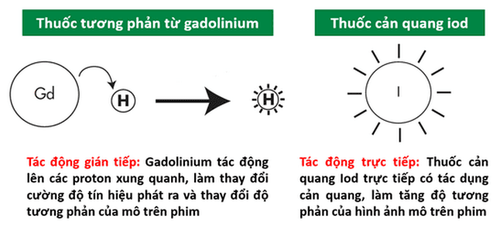
Magnetic resonance imaging (MRI) is a noninvasive medical test that doctors use to diagnose and treat medical conditions. MRI does not use ionizing radiation (X-rays) at all, like X-rays or computed tomography scans, so there is no risk of exposure.The principle of a specialized joint mri scan is to use radio frequency, magnetic energy and a computer to create images of the bones, muscles, tendons, ligaments and cartilage in the knee and the surrounding area at any joint position. Together with the participation of contrast agents from injection into the blood vessel lumen, the drug will help the pathophysiological phases on the lesions to be shown more clearly.
MRI tests can be tailored to specific joints. Of these, large joints such as shoulders, hips, knees, ankles and feet are most often concerned due to the frequency of common diseases, but basically all joints can be evaluated. The advantage of MRI is that it helps to show joint anatomy to a higher degree than other imaging modalities such as x-rays or CT. Detailed MRI images of the joints allow doctors to determine the presence of a number of conditions. Common indications include evaluation for causes of pain such as from trauma, osteoarthritis, or osteoarthritis. MRI is also very good at evaluating soft tissues around joints that can become inflamed, like tennis elbow.)
In addition, other conditions that may be diagnosed with a joint MRI include joint tumors, arthritis, genetic abnormalities, and bone and joint disease. Usually, when approaching patients with joint problems, the first indication is musculoskeletal ultrasound or joint X-ray. However, with magnetic resonance imaging of the joints with injection of magnetic contrast, which is a semi-invasive means, the structural components at the joint will show up clearly, helping the doctor to identify the damage and adjust. diagnose.
2. Indications when needing magnetic resonance imaging of joints with injection of magnetic contrast agent
Imaging acquired with contrast-enhanced magnetic resonance imaging is necessary in the following situations:Assess for any damage within the joint, eg tear of the anterior cruciate ligament at the knee joint , and whether it causes inflammation Assess for degenerative joint wear and tear such as cartilage loss or rupture of the articular capsule causing inflammation due to osteoarthritis Assess for any inflammation in the joint and of surrounding structures such as tendons, ligaments, bursitis Assess the reason for local swelling or pain in relation to the joint Assess the nature of the tumor: location, size, enhancement

Hình ảnh sau khi chụp cộng hưởng từ khớp có tiêm thuốc đối quang từ
3. Joint magnetic resonance imaging procedure with injection of magnetic contrast agent
Because the presence of metal is a contraindication to MRI scans in general, it is important to let your doctor know if you have a pacemaker, metal foreign bodies such as shrapnel, or other types of debris. clip, clamp aneurysm bag.You can wear your personal clothes normally as long as they do not contain any metal components, including buttons, zippers and metal threads. However, to ensure the most ideal shooting conditions, you should change into specialized clothes for magnetic resonance imaging at the imaging room; at the same time, this also makes you more comfortable while lying still for a long time. In addition, you will also be asked to remove all jewelry, glasses, dentures, wigs, wallets, bank or credit cards... from your body.
Because you have magnetic resonance imaging with magnetic contrast injection, you need to fast the night before the scan, except for medicine for chronic diseases or pain medicine for acute joint disease to help Magnetic resonance imaging becomes more comfortable, as you will have to lie still during this time.
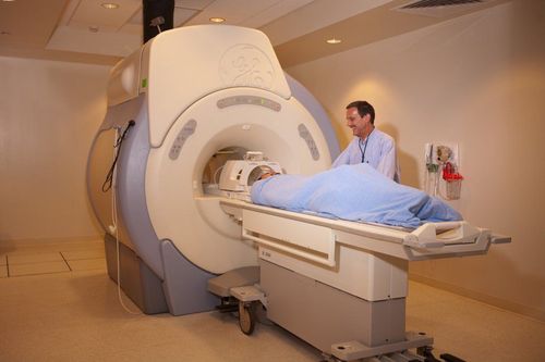
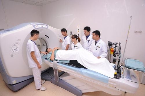
When the preparation for magnetic resonance imaging of the joint with contrast injection is ready, a slide in the machine's lap will come out. You need to lie on a table, with your head or legs in front, depending on the type of joint to be assessed as an upper or lower body joint. The slide will then move, bringing the scanned body into the center of the body, which is essentially a giant magnet tube. This whole process will be observed by the control technicians in the next room. Once the area to be surveyed has been located, the technician will operate the machine. You will hear a very loud noise while the machine is scanning. Therefore, if the noise is too loud, you will be able to wear earplugs or headphones to listen to your favorite music and relax during the shooting process.
If you have a feeling of suffocation, anxiety, please inform the technician to prescribe medicines that can help you feel more comfortable. However, if you need this medicine, you will need to have a loved one drive you home after the scan and you will also need to avoid operating a vehicle or machinery for at least one day afterward.
The first phase is a joint magnetic resonance imaging phase without contrast injection usually within 30 to 45 minutes. If only one joint is examined, the time will be shortened. When ensuring that the collected images are of good quality, the technician will inject contrast material into the vessel and perform the same image collection when estimating the time it takes for the drug to reach the joint.
When the scan is over, the technician helps you to sit up and takes you out. At this point, you can go home and do your normal activities as your bones and joints allow.
4. What to monitor after magnetic resonance imaging with magnetic contrast injection?
The time required to read the results is often longer and requires a specialist radiologist to evaluate and analyze. Results will be sent back to the appointing doctor. By combining the medical history, symptoms, physical examination, and other imaging findings, the physician will make a diagnosis and guide treatment.In case of joint damage requiring surgical intervention, the results of joint magnetic resonance with injection of this contrast agent can be the basis to help shape the surgical protocol to be performed as well as to provide evidence for the compare and contrast the effectiveness of treatment in the future.
Compared with magnetic resonance imaging of joints without injecting contrast, drug injection does not require any special monitoring. The observed rate of allergic reactions due to contrast is very low, much lower than when contrast is used in computed tomography. However, a very small number of patients may experience pain at the injection site or a bitter, metallic taste in the mouth a few days later. Even so, these manifestations are transient and do not cause significant effects.
In summary, joint magnetic resonance with injection of magnetic contrast is the best means to help identify joint damage. With the role of contrast agent, the anatomical structures will be clearly shown, helping the doctor to identify joint damage in the best conditions and offer effective intervention directions, improving as well as preserving function. long-term joint mobility.
Vinmec International General Hospital is one of the hospitals that not only ensures professional quality with a team of leading medical doctors, modern equipment and technology, but also stands out for its examination and consultation services. comprehensive and professional medical consultation and treatment; civilized, polite, safe and sterile medical examination and treatment space.
Doctor Giang has many years of experience in the field of diagnostic imaging, especially in the field of multi-slice computed tomography, magnetic resonance. Currently, the doctor is working at the Department of Diagnostic Imaging and Nuclear Medicine - Vinmec Times City International General Hospital.
For medical examination and treatment at Vinmec, please go directly to Vinmec medical system nationwide or register online HERE.