This is an automatically translated article.
Article by Doctor Pham Quoc Thanh - Department of Diagnostic Imaging - Vinmec Hai Phong International General Hospital
Cranial X-ray is an imaging technique performed under an x-ray machine and is of high value in detecting and diagnosing diseases related to the bones of the skull (such as tumors, trauma, etc.) injuries, diseases of the sinuses,...) and the brain (increased intracranial pressure, brain tumor).
1. What is a cranial X-ray?
The brain is the control center of the central nervous system, responsible for controlling behavior, and is a particularly important organ that coordinates the activities of all organs and parts of the human body. The skull is a hard shell of bone that surrounds and protects the brain. Any change in the structure of the skull bones can lead to brain damage and conversely, brain parenchymal diseases can also lead to changes in skull bone morphology.Cranial X-ray includes 2 normal upright and inclined positions, in addition to other special positions such as Blondeau, Hirtz, Schuller, Stenvers...
2. When is a brain X-ray indicated?
Patients with suspicion of falling into one of the following cases will be assigned to have a skull X-ray:
Diseases of the skull bones such as:
Trauma, traumatic brain injury Cranial malformations Inflammation of the sinuses U skull bones Intracranial diseases such as:
Increased intracranial pressure. Brain tumor: meningioma, pituitary tumor...
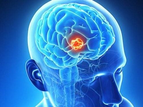
Người có bệnh lý u não sẽ được chỉ định chụp Xquang sọ não
3. Instructions for reading straight and lateral cranial X-ray films
3.1. Based on the shape of the skull dome Normal image:
The skull is composed of 2 thin, flattened bones called the inner and outer cranial plates, in between the two bones is spongy bone containing veins. The length and height of the skull are proportional to each other. Abnormal image:
Abnormal image of skull shape. There may be subsidence, skull fracture due to trauma or tumor destruction. 3.2. Based on the joint lines of the skull bones Normal picture:
The junction between the bones (frontal bone, parietal bone, temporal bone, occipital bone, scapula) is called cranial sutures. Clinically, there are 2 important joints including: parietal frontal joint and parietal occipital joint. + In children, because the joints of the skull are not fully developed and closed, they form fonts including anterior fontanelle, anterior fontanelle, posterior fontanelle, and posterior fontanelle.
+ In an adult, the skull bones are in close contact with each other and the skull joints are serrated,.
Abnormal picture:
If the bones do not come into contact with each other and the skull joints are dilated, it may be a sign of increased intracranial pressure. At the same time, skull joint dilation is also one of the signs of traumatic brain injury.
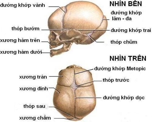
Đường khớp xương sọ
3.3. Based on the pituitary To be able to observe and comment on the pituitary, it is necessary to rely on the X-ray film of the brain in the inclined position.
Normal picture:
The pituitary fossa has a structure consisting of the anterior and posterior ganglia, the lumen of the fossa and the crater. Abnormal picture:
If there are related pathologies, there may be some changes in the pituitary fossa as follows:
The anterior saddle is attached to the posterior node due to calcification of the intertrochanteric ligament. The pituitary fossa is enlarged (spreading into a concave shape), dilating the pituitary fossa caused by a tumor. Widen the mouth of the saddle.
3.4. Based on the dot impression, the picture is normal:
On the cranial X-ray film, you can observe the images like finger prints, this is the point impression. In normal people, the imprint usually only begins to appear at the age of 8, most clearly between the ages of 20 and 25, and as age increases, it becomes less and less obvious. If observed on the cranial X-ray film in the upright, inclined position, the imprints will be clearly indicated in the temporal region. Abnormal imaging:
Marking points only in patients with increased intracranial pressure.
3.5. Based on the vascular lines On the normal craniogram, intracranial blood vessels will be seen - small, branched bright lines. Especially in the elderly, the image of veins will be clearly seen in the skull film.
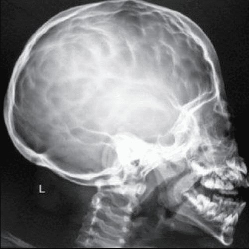
Hình dấu ấn ngón tay trong tăng áp lực nội sọ
4. Brain X-ray at Vinmec Hai Phong International General Hospital
Vinmec Hai Phong International General Hospital is currently one of the hospitals in the Vinmec international hospital system equipped with modern machinery and equipment to serve the medical examination and treatment process.
Patients who have X-ray of the brain at Vinmec Hai Phong International General Hospital will be clearly guided by the support staff and technical staff on the manipulations and procedures to minimize exposure. X-ray exposure to reduce the impact on the health of the photographer, ensuring the best diagnostic image.
If your X-ray image has any abnormality, the doctors will consult promptly and offer an effective treatment plan.
To register for examination and treatment at Vinmec International General Hospital, you can contact the nationwide Vinmec Health System Hotline, or register online HERE.


