This is an automatically translated article.
The article is expertly consulted by Master, Doctor Nguyen Hong Hai - Doctor of Radiology - Department of Diagnostic Imaging and Nuclear Medicine - Vinmec Times City International General Hospital.Computed tomography of the thoracic spine with contrast injection is a widely used technique to diagnose trauma, tumors, and inflammation of the bones and soft tissues of the thoracic spine.
1. Computed tomography of the thoracic spine with contrast injection is indicated in which cases?
Computed tomography (CT) scan is a technique that uses X-rays to scan the area of the body that needs to be examined. The tomography machine is linked to the computer system, the image after being taken will be processed by the software to create clear 2D or 3D images. These images help the doctor accurately assess the patient's condition. The method of computerized tomography has a fast analysis and image processing speed, so it is often used in emergency cases. Because of its many advantages, computed tomography is being widely used in many fields, in addition to imaging diagnosis, also used in surgical guidance, radiation therapy, postoperative monitoring,... To image The image obtained is clear, with better contrast, in some necessary cases, the doctor may prescribe contrast injection when taking pictures.The thoracic spine consists of 12 vertebrae, intermediate in size between the cervical vertebrae and the lumbar vertebrae. The thoracic vertebrae work with the ribs and sternum to form the ribcage. Computed tomography of the thoracic spine is a commonly used technique today to evaluate lesions of the thoracic spine, discs, thoracic spinal canal and adjacent components. When contrast is injected, it helps to detect and evaluate diseases such as inflammation, tuberculosis, tumors of the spine, spinal cord, osteomyelitis and soft tissue of the thoracic spine,
Computed tomography of the thoracic spine with injection Contrast has no absolute contraindications. However, doctors will be especially careful and consider before performing for pregnant women, patients with renal failure, and people allergic to iodinated contrast agents.

Tiêm thuốc cản quang khi chụp MRI sẽ cho hình ảnh rõ nét hơn
2. To perform computed tomography of the thoracic spine with contrast injection, what preparation is required?
The implementation team includes specialist doctors, radiology technicians, nurses. The necessary facilities include:Computer tomography machine; Specialized pumps; Film, cassette, image storage system. In addition, to perform contrast injection, you need supplies and drugs such as: syringe, needle, cotton, gauze, skin antiseptic solution, iodinated contrast soluble in water, distilled water or saline physiology, medicine box and emergency equipment for contrast agents,...
Before performing the technique, the patient will be carefully explained by the medical staff about the purpose of the scan, how to take it, and how to lie down. , how to hold your breath, ... to coordinate with the doctor. Patients should remove jewelry such as earrings, necklaces, hairpins and other metal objects if present. The medical staff will tell the patient to fast, drink at least 4 hours before the procedure, can drink water but not more than 40ml. The doctor may prescribe a sedative to help relax if the patient is excited, not lying still.

Bệnh nhân sử dụng thuốc an thần nếu bị kích động
3. Steps to conduct a CT scan of the thoracic spine with contrast injection
3.1. Patient's pose
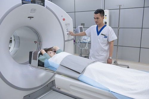
The performance team helps to place the patient in the machine frame, lying on his back, with the shoulders lowered as much as possible, with both hands raised up along the body axis. To get a clear image, the patient needs to hold their breath and not swallow during the scan.3.2. Steps to conduct the technique of computed tomography of the thoracic spine with contrast injection
The thoracic spine is localized in two planes. Take the lateral position image (sagital) starting from the upper border C7 to the lower L1. Depending on the clinical requirements, the doctor will install the appropriate scan program. To evaluate the pathology of herniated discs can use the cut in the direction of the discs or take the entire thoracic spine. After capturing, use image processing software. Select film images on bone windows, disc windows. Cut again after contrast injection. When taking a CT scan of the thoracic spine, if the patient is not lying motionless, the resulting image will be blurry and noisy and need to be re-imaged.
Hình ảnh chụp MRI cột sống ngực được xử lý qua phần mềm
4. Assess the results of computed tomography of the thoracic spine with contrast injection
Evaluation of vertebral body injuries such as: vertebral body rupture, vertebral body collapse, vertebral body slip, displacement of posterior vertebral wall injury, posterior arch lesions, traumatic hematoma, paravertebral soft tissue injury, Contrast foreign body... Injuries in degenerative spondylosis such as: degenerative bone spurs, ligament calcifications, lateral mastoid hypertrophy, spinal stenosis, spondylolisthesis due to waist opening... assessment of congenital abnormalities of the spine: congenital scoliosis, spina bifida... Tumors of the vertebral body, in the spinal canal, and in the paraspinal software Compare the images before and after injection medications to identify and evaluate comorbidities.
Mẩn ngứa là một dị ứng có thể xảy ra với người bệnh khi tiêm thuốc cản quang
5. Complications and treatment
CT scan of the thoracic spine with contrast injection is a safe technique with few complications. Complications, if any, often occur because the patient is allergic to the contrast agent. Therefore, after the injection, the patient will be closely monitored, if there are signs of an allergic reaction to the contrast agent such as nausea, dizziness, rash, itching, ... the technical team will handled according to the process of diagnosis and treatment of contrast agent accidents according to regulations of the Ministry of Health.In general, CT scan of the thoracic spine with contrast injection is a modern and advanced imaging technique that plays an important role in diagnosing diseases of the spine, discs, spinal canal and other components. neighboring part.
In order to meet the needs of medical examination and treatment, currently Vinmec International General Hospital has been and continues to bring in a system of modern machines such as magnetic resonance imaging (MRI), computed tomography (CT), X-ray, etc. ... in the work of medical examination and treatment, diagnostic imaging, disease treatment. Especially in order to bring high efficiency in medical examination and treatment, Vinmec now also designs many accompanying medical services, bringing many conveniences to customers.
Master, Doctor Nguyen Hong Hai graduated with a Master's degree in Diagnostic Imaging at Hanoi Medical University with strengths in diagnosing breast and thyroid cancer. Currently, Dr. Hai is working at the Department of Diagnostic Imaging, Vinmec Times City International Hospital.
You can directly go to Vinmec Health System nationwide to visit or contact the regional hospital hotline here for support.


