This is an automatically translated article.
Radical lymphadenectomy is often used to treat cancer with metastatic enlarged cervical lymph nodes. When a cervical lymphadenectomy is indicated, the patient should absolutely follow all instructions given by the doctor.1. What is cervical lymphadenectomy?
Radical cervical lymphadenectomy (traditional classical) is a surgical method to remove lymph nodes at the angle of the jaw, the clavicle, the lateral margin of the sternohyoid muscle, the hyoid bone, the anterior abdomen of the biceps muscle, and the anterior margin of the quadriceps muscle. ladder (levels I - V) and many important anatomical components such as internal jugular vein, sternocleidomastoid muscle, and XI cord on one side of the neck.
When performing radical cervical lymphadenectomy, the doctor needs to preserve the carotid artery, sympathetic nerves, XI, diaphragmatic, XII, and chin branches of VII.
Radical cervical lymphadenectomy is not removal of parotid gland, occipital region, lateral - posterior pharyngeal space, anterior vertebral column. Doctors usually perform cervical lymphadenectomy before removing the primary tumor in the ENT region and head and neck in the same surgical anesthesia.
Bác sĩ thường thực hiện nạo vét hạch cổ trước cắt bỏ khối u nguyên phát trong cùng 1 lần gây mê phẫu thuật.
2. Indications and contraindications for radical cervical lymphadenectomy
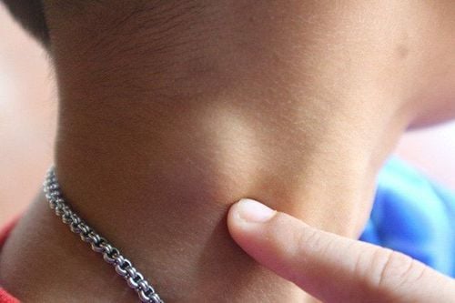
2.1 Indications Radical cervical lymphadenectomy is indicated for cases of lymph node metastasis, cervical lymphadenopathy over 3cm (N2a, N2b, N3), including primary metastatic cervical lymph nodes. The most common types of cervical lymph node metastasis are cancers of the gastrointestinal tract - upper respiratory tract.
2.2 Contraindications There is no absolute contraindication for radical cervical lymphadenectomy, except in cases where the cervical lymph node has been attached to the carotid artery. Cervical lymphadenectomy should be used with caution in patients who are elderly (over 70 years of age) or have high blood pressure and diabetes.
3. The procedure of dredging the cervical lymph nodes
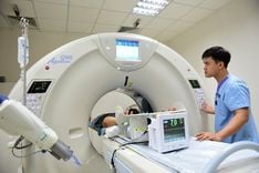
3.1 Prepare Personnel to perform: Doctors, nurses, technicians proficient in head and neck cancer surgery. The operating team consists of 1 main surgeon, 1 auxiliary surgeon and 1 instrument technician; Technical means: Surgical kit for head and neck area, need to use more electric knife, bipolar electrocoagulation; Patient: To be explained about the purpose of surgery and the risk of possible complications, consent to perform surgery; Medical records: Prepare all kinds of documents as prescribed. In addition, results of neck ultrasound and CT scan are required to assess the extent of lymph node infiltration and carotid adhesions. 3.2 Performing procedures Examination of records: Including examination of tests, medical records,...; Examination of the patient; Stage 1: U-shaped skin incision between the mastoid process on both sides if both larynx and hypopharynx are removed. Or make an L-line incision; Stage 2: Anterior limited dissection to free the subungual area. In this step, the doctor goes from the midline white line at the level of the thyroid ring to the nail, exposing the inferior border of the biceps muscle to the angle of the jaw. Then, the doctor removes the connective tissue - lymph nodes in the anterior area of the hyoid bone. The doctor often ligation, remove the anterior jugular vein in order to remove the connective tissue in the patient's lower parotid gland. Simultaneously, dissection revealed facial veins and veins (can be ligated), dissected and preserved the tongue and XII cords, and exposed the body of the facetilla. The anterior limit is removal of all connective tissue - subungual, supranadal, submandibular, and parotid glands; Stage 3: Dissection of the biceps carotid region and the XI cord. The surgeon made an incision in the area below the mastoid process, removing the head above the sternocleidomastoid muscle. The surgeon then dissects the fatty layer to find the XI cord and the internal jugular vein, which are separate from the X cord and the internal carotid artery. The doctor will remove the connective tissue - the carotid biceps ganglion and the XI cord, tie the head on the internal jugular vein, cut off the head above the sternocleidomastoid muscle and dissect the submandibular area; Stage 4: Anatomy of the jaw angle: From the 2nd and 3rd stages, the biceps, carotid aneurysms, and XII cords were revealed, allowing ligation and cutting of small vessels such as the superior sternoclavicular artery, the occipital artery. , the body of the face and tongue, dissecting the carotid aneurysm, along the carotid bundle descending to ligate, remove the pharyngeal venous plexus and also the thyroid gland. Here, the doctor dredged the anterior triangle of the neck and the biceps carotid region, removed connective tissue, fat, and lymph nodes from the posterior biceps carotid region, anterior submandibular region, from the posterior biceps subabdominal, From top to bottom, it runs along the axis of the internal jugular vein and also the vein, the sternocleidomastoid muscle. From there, it is allowed to continue dissection below the supraclavicular region; Then 5: Dissect the area above the clavicle. The doctor performed a scapular hysterectomy, continued dissecting downwards to separate the internal jugular vein from the X cord and internal carotid artery, and the filter behind was the vein and transverse cervical artery. The surgical site allows the excision, dissection, and removal of connective tissue, fat, and supraclavicular lymph nodes. From here, the surgeon can dissect the head below the sternocleidomastoid muscle to remove the same inferior end of the internal jugular vein to remove the radical cervical lymph node. The doctor should pay attention to the removal of the internal jugular vein, which needs to be clamped with surgical forceps without clip, after ligation, it is necessary to sew the end with Vicryl thread. After dissection and removal of the lymph node group from I-V, four anatomical landmarks in front and bottom are the trachea, above and below the abdomen, behind the biceps muscle, above and behind the transverse process of the cervical vertebrae, behind and below is the brachial plexus; Stage 6: Restoration of the surgical cavity. The doctor performed a thorough hemostasis check, placed a closed drain, closed the surgical cavity with 2 planes of skin and skin attachment, and then applied pressure to the neck area with moderate tightness. 3.3 Follow-up after intervention Bleeding: If there is pink fluid in the drainage vessel after surgery, the amount of fluid gradually decreases, after 36 or 48 hours, the drainage can be withdrawn. If the patient is bleeding, the drain will be full of bright red blood, pulse, blood pressure drops, the patient is slow to wake up and panicky; Lymphatic leak: The patient has a white discharge like rice water when draining to the bottle, which can leak up to 500ml/day; Shoulder pain: Usually occurs in cases of removal of the XI cord.

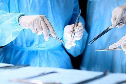
Sau khi thực hiện phẫu thuật nạo vét hạch cổ tiệt căn, người bệnh phải tuân thủ lời khuyên của bác sĩ và theo dõi chặt chẽ sức khỏe của mình
3.4 Risks and management measures Bleeding: Physicians need to prevent the risk of bleeding after radical cervical lymph node dissection by paying attention to skin margins, small venous knots, and internal jugular vein closure. ; Lymphatic leak: If after changing the compression bandage, the patient continues to have a large amount of white discharge, the doctor needs to open the surgical cavity to check the chest tube area, clamp and tie: Shoulder pain: The external branch of the XI nerve can be preserved, if this condition persists after surgery, then it can be treated with physical therapy. Radical lymph node dissection may have some undesirable risks. Therefore, to ensure safety, patients should follow the doctor's advice and closely monitor their health after the procedure. To treat head and neck cancer with the most effective radical cervical lymphadenectomy, patients should choose reputable and quality medical centers. Vinmec International General Hospital gathers a team of famous doctors and nurses in the industry, with good expertise, rich experience, heart and vision. Not only domestic doctors, there are also foreign doctors from Japan, Singapore... helping patients access new and effective treatment regimens from countries with developed medical backgrounds. In the world.
Please dial HOTLINE for more information or register for an appointment HERE. Download MyVinmec app to make appointments faster and to manage your bookings easily.