This is an automatically translated article.
The article was written by Master, Doctor Nguyen Van Khanh - Pathologist - Laboratory Department - Vinmec Times City International General HospitalStandard procedures and methods are used to process nearly all types of biopsies. These procedures are the usual ways in which patient samples are prepared in the laboratory. Other procedures, described below, may also be performed on certain types of specimens (eg, lymph nodes or bone marrow).
1. How are biopsy specimens routinely handled?
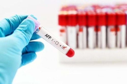
After being removed from the body, the biopsy specimen is placed in a vial containing formalin or some other liquid to preserve the sample. The sample vial is labeled with the patient's name and other identifying information (such as the patient's medical record number and date of birth) and the biopsy site (exactly where on the body the sample was taken). specimens are removed). It is then sent to the laboratory. Next, a pathologist or trained laboratory staff member visually observes the specimen. This process is called a gross examination. This is done to record what is seen by looking at, measuring, or touching a tissue sample. Macroscopic examination includes measuring the size, characterization, color, density, and other characteristics of the tissue sample. The laboratory staff may take pictures of the tissue sample as part of the record. Macroscopic examination is very important because the pathologist can see features that are suggestive of a certain type of cancer. It also helps the pathologist decide which parts of a large biopsy are most important to take for microscopic examination.
For all minor biopsies, eg with perforation or core biopsies, the entire specimen is usually observed under the microscope. The tissue sample is placed in a small container called a cassette. The cassettes are used to hold the tissue sample carefully during sample handling (tissue transfer). Tissue transfer can take several hours (but is usually done overnight). After the tissue transfer, the specimen is placed in a hot paraffin wax mold. The wax coagulates to form a solid mass that protects the tissue sample.
This tissue sample molded in paraffin wax is cut into very thin slices using an instrument called a microtome. Thin slices of this tissue sample are placed on slides and embedded in a series of dyes that change the color of the tissue sample. The color makes the cells easy to see under the microscope. For most biopsies, this routine processing is all that is needed. At this point (usually a day after the biopsy is done), the pathologist will look at the tissue sample under a microscope to make a diagnosis. Observation of solid specimens processed in this way is called histology, which is the study of the structure of cells and tissues.
2. Special biopsy sample handling: Cold biopsy, also known as instant biopsy (intraoperative consultation)
Some tissue sample information is essential during surgery to make immediate decisions. If it is not possible to wait a few days for the results of routine histopathology, the surgeon will recommend a pathology consultation during surgery. Such surgical consultation is often referred to as cryo-biopsy or immediate biopsy.
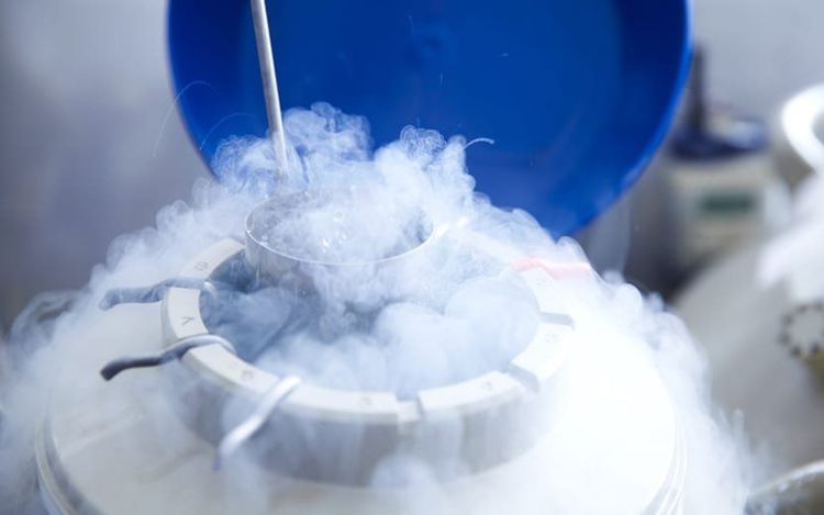
2.1 So how is a cold biopsy done?? Fresh tissue samples (not soaked in any solution) after being removed from the patient's body must be sent from the operating room immediately to the pathologist. The pathologist will perform a macroscopic examination and decide which part of the sample to take for microscopic examination. Instead of treating the tissue sample in waxy clumps as is the case with routine testing, in a cryo-biopsy the tissue sample is rapidly frozen in a special solution, forming an ice block around the tissue sample. It is then thinly sliced on a special device, then rapidly stained (dipped in a dye), and finally observed under a microscope. Cold biopsies are often not as revealing of tissue features as those obtained from tissue samples cast in wax, but they are good enough to help the surgeon make further management decisions during surgery. surgery for the patient.
2.2 When is a cold biopsy done? To find out if a tumor is cancerous: Sometimes the type of surgery depends on whether the tumor is cancerous (malignant). For example, surgery to remove a tumor may be enough to treat it if the tumor is not cancerous (benign), but if the tumor is cancerous, surgery may need to remove more tissue. and/or accompanying lymph nodes. In such cases, the surgeon must send the tumor for cryopreservation. This test may provide enough information to help surgeons decide which surgical approach is best for the patient. However, sometimes a cold biopsy doesn't give a definite answer and the piece of tissue will need to go through routine testing or even special handling to get a definite answer. If this is the case, the surgeon usually stops the operation and closes the incision. Once the results are available, another surgery may be needed.
To make sure all the cancer is removed: Cancer surgery often strikes a balance between removing all of the cancer with minimal damage to surrounding normal tissue. If the surgeon is concerned that the cancer hasn't been completely removed, a slice from the margin of the removed tissue (also called a section) will be sent for a cryo-biopsy. If the cut area is free of cancer cells, no further resection is usually needed. But if the cut area still contains cancer cells, then some cancer cells can be assumed in the patient's tissue. In this case, the surgeon will remove more tissue to try to remove all the cancer cells and reduce the chance for the cancer cells to come back. If the surgeon cannot remove more tissue, there may be other treatment options, such as radiation therapy to kill any remaining cancer cells.
Mohs procedure (microscopically controlled surgery)
This procedure is used to treat certain types of skin cancer. In Mohs surgery, the surgeon removes a thin layer of skin that the tumor can invade and then examines the specimen under a microscope. If cancer cells are seen, more layers are removed and examined until no more cancer cells are seen on the skin specimens. The process is slow but means it can save more of the normal skin near the tumor. Mohs surgery is a highly specialized technique that can only be performed by trained doctors.
3. How are cytological specimens handled?
How cytology specimens are handled depends on the type of specimen. Some patient samples are smeared on slides by the physician collecting the sample. These slides, called slides, are sent to a cytology laboratory where they are dipped in a series of colored dye solutions, like those used for biopsies.
Other specimens, such as body fluids, cannot be spread easily on slides because they are too dilute (too few cells in a large volume of liquid). They will be centrifuged to gather these cells onto a slide before they are stained.
After being treated and stained, the samples are examined under a microscope. Abnormal cells are found and marked with a special pen. The pathologist will then examine the marked cells to make a diagnosis.
4. How long are patient samples stored?
Each country has a regulation that requires laboratories to store samples for a certain period of time. For example, in the United States, federal law requires laboratories to keep:
Cytological specimens for at least 5 years. Histopathological specimens are at least 10 years old. Paraffin wax blocks at least 2 years. At the Pathology Unit - Vinmec Times City Hospital. The cytological records are stored for at least 5 years. Histopathological specimens and paraffin wax blocks have been stored since the establishment of the hospital (nearly 8 years) and are kept for at least 10 years.
What this means for you
Some people like to get a second diagnostic view from their tissue samples (patient samples). This is called a reevaluation of the results. It means having another doctor look at your biopsy tissue and make a diagnosis based on what that doctor sees.
Human tissue samples are not removed immediately after testing. So, in most cases, if enough tissue is available, the sample can be sent to a doctor or another laboratory. Sometimes storing tissue samples for a longer period of time can be helpful in other ways. For example, someone with cancer who had their first tumor removed, a few years later they have another tumor, and now the doctors want to know if this new tumor is an old tumor that has come back (recurred). or a completely new tumor. This can be differentiated based on the location of the two tumors and by comparing histopathological specimens from both specimens. But sometimes, more tests (such as immunohistochemistry) that can be done using tissue from the paraffin wax block of the original tissue sample are also helpful.
Vinmec International General Hospital is one of the hospitals that not only ensures professional quality with a team of leading medical doctors, modern equipment and technology, but also stands out for its examination and consultation services. comprehensive and professional medical consultation and treatment; civilized, polite, safe and sterile medical examination and treatment space.
Any questions that need to be answered by a specialist doctor as well as customers wishing to be examined and treated at Vinmec International General Hospital, please register for an online examination on the Website for the best service.
Please dial HOTLINE for more information or register for an appointment HERE. Download MyVinmec app to make appointments faster and to manage your bookings easily.


