This is an automatically translated article.
The article is professionally consulted by Master, Doctor Nguyen Van Huong - Department of Diagnostic Imaging - Vinmec International Hospital Da Nang. The doctor has many years of experience in the field of diagnostic imaging.Kidney disease is one of the diseases that can leave serious complications if not examined and treated promptly. Currently, ultrasound diagnosis of pyelonephritis is often prescribed by doctors to identify and properly assess the disease condition, then provide an effective treatment plan.
1. Is pyelonephritis dangerous?
Renal pyelonephritis is the dilation of the calyx and renal pelvis due to fluid retention in the kidneys. If the kidney is dilated for a long time, it will deform, swell, and thin, like a balloon filled with water and can burst at any time. Renal calyx disease can occur at any age, depending on the different degree that the sufferer shows the following symptoms:Usually, people with a history of kidney disease have pain in the hip area ( 1 or 2 sides). Pain in the lower back, ribs, can spread to the lower abdomen. Frequent urination, urinary incontinence, painful urination, and pain. Hematuria . Fever, headache, dizziness, nausea Sudden increase in blood pressure Accordingly, the most common cause of pyelonephritis is urinary stones, especially when kidney stones fall into the ureter. obstruction here, causing fluid retention in the entire area from the ureter. In addition, prostate enlargement, kidney tumor, ureteral tumor, ureteral scarring, ureteral foramen, narrowing of the bladder neck... can also cause obstruction and cause dilatation of the renal calyces.

2. The procedure of performing ultrasound calyx dilatation of the renal pelvis
Ultrasound diagnosis of pyelonephritis is a technique that helps doctors early detect and accurately diagnose the cause of the disease. Accordingly, the procedure of performing pyelonephritis ultrasound is carried out in accordance with regulations, based on the position of the kidney in the body, the doctor can choose the best cross section during the ultrasound to get the image. vertical and horizontal of the kidney.Renal ultrasound in adults gives normal results when the images have the following features:
Kidneys are shaped like a pea, with a navel in the middle. The size of the two kidneys is often not the same; The border is regular, the left side has splenic pressure, so the left kidney parenchyma is triangular in shape. The renal cortex has a lower negative density than the liver and spleen. The renal pyramid has a slightly lower acoustic density than the cortical part. The part of the renal sinus complex has the greatest negative density. The collecting system (pyelonephritis, calyces, and calyces) is normally a narrow, inconspicuous urine-filled cavity that is clearly seen as a negative void in a well-diuretic person. The renal pelvis can also range from a small acoustic drum structure within the kidney to a large acoustic drum protruding from the kidney. Normal renal ultrasound images in children are different from those of adults as follows:
The glomerulus is more concentrated in the cortex, so the sound density is higher than that of the adult kidney. The kidneys in young children have very little fat, so the renal sinus complex contains only structures of the calyx system. The calyx system is relatively stretched at about 75%, so the calyx and calyx are fluid-containing structures.
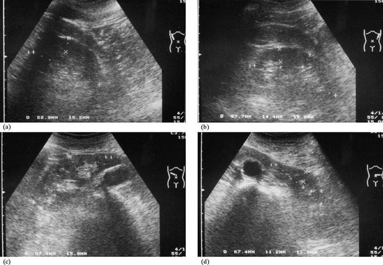
Grade 1: the renal pelvis and the renal calyces are slightly dilated. Grade 2: The renal pelvis and renal calyces are markedly dilated, compressing the renal parenchyma to narrow. Grade 3: renal pelvis and calyx dilate into a large cyst, indistinguishable structures of the collecting system. Renal parenchyma is very thin. In addition, ultrasonography of pyelonephritis can also see the location of ureteral stones, congenital ureteral atrophy, renal tuberculosis and other causes of renal calyces.
3. Is renal calyces dangerous?
After having ultrasound results to diagnose pyelonephritis, the doctor will evaluate the extent and ability to recover according to each stage of the disease.1st and 2nd degree pyelonephritis: In 1st and 2nd degree, the damage is not too serious, it is still possible to recover. Grade 3 pyelonephritis: Level 3 is a particularly dangerous level, the renal wall is dilated and thin, losing its elasticity, the risk of complications is very high, requiring early treatment. However, in the early stages of pyelonephritis, there are often no symptoms, so it is often ignored by patients. Therefore, regular health check-ups to detect diseases early is very necessary to avoid dangerous complications that may occur.
Some dangerous complications of pyelonephritis such as:
Kidney infection: If urine accumulates for a long time in the kidney, it will create conditions for bacteria to grow causing inflammation, interstitial infections, renal calyces.. These diseases often recur many times, seriously affecting kidney function.
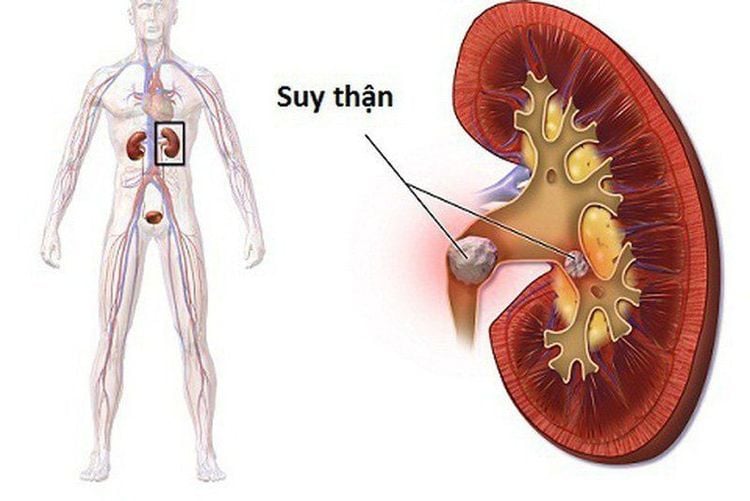
4. How to treat pyelonephritis?
Kidney disease is a dangerous disease, so early examination and treatment will give a high chance of treatment. However, treatment depends on the cause and extent of the disease. The goal of treatment is to reduce the pressure on the kidneys, preserve better kidney function, and eliminate the cause of the disease. Some treatment methods for pyelonephritis are as follows:4.1. Urinary catheterization
In most cases of pyelonephritis due to hydronephrosis, the first treatment is to insert a catheter into the ureter to create a flow that allows urine to flow, reducing pressure on the kidneys. A ureteral catheter may be inserted through the urethra or placed downstream through the kidney through a small incision in the skin to enter the renal pelvis.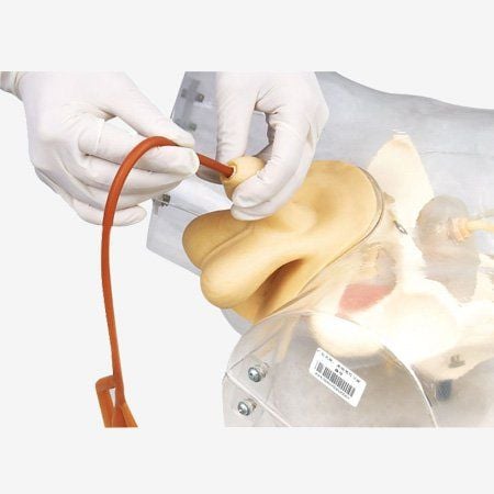
4.2. Treat the cause of the disease
Once the pressure on the kidney has been relieved, it is time to rely on the cause of the obstruction to treat. Usually due to kidney stones, urinary stones, doctors will use some drugs such as: ureteral smooth muscle relaxants, anti-inflammatory drugs, pain relievers, antibiotics, diuretics, potassium citrate salts, drugs. reduce uric acid levels... to help patients reduce symptoms, facilitate stone elimination. Or percutaneous lithotripsy, endoscopic retrograde lithotripsy, ... However, the doctor will prescribe treatment or not depends on the cause of the dilation, kidney function and clinical symptoms. of the patient through the image of renal pelvis dilatation on ultrasound.In addition to complying with the treatment prescribed by the doctor, the patient also needs to build a suitable nutrition and scientific lifestyle, drink lots of water and increase daily exercise. Kidney disease is a dangerous disease that, if not detected early, can have serious consequences for health. Therefore, performing ultrasound and diagnosis when there are symptoms of disease is extremely necessary.
Currently, Vinmec International General Hospital has full facilities, modern ultrasound machines and other advanced methods to help find the cause of dilation to have appropriate treatment such as: CT scan 460 slices cutting, MRI 3.0Tesla. Accordingly, the entire procedure of performing pyelonephritis ultrasound is well-trained and experienced by a team of medical professionals, with many years of experience, so the patient can rest assured with the results. diagnostic results. When the results are accurate, the patient will be consulted for treatment, ensuring the best health.
Please dial HOTLINE for more information or register for an appointment HERE. Download MyVinmec app to make appointments faster and to manage your bookings easily.


