This is an automatically translated article.
The article was professionally consulted by Specialist Doctor I Tran Cong Trinh - Radiologist - Radiology Department - Vinmec Central Park International General Hospital. The doctor has many years of experience in the field of diagnostic imaging.Pericardial calcification is one of the common problems in patients with constrictive pericarditis. Early diagnosis of pericardial calcifications through methods such as ultrasound, X-ray,... brings great value for effective disease treatment.
1. What is pericardial calcification?
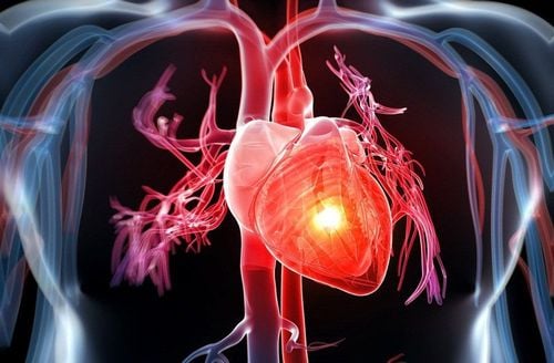
1.1 Anatomy and physiology of the pericardium The pericardium covers the heart and the proximal portion of the great arteries and veins that arise from the heart. Pericardium consists of parietal and visceral leaves. The pericardium is attached to organs including the spine, sternum, and diaphragm by ligaments. This organ is regulated and nourished by lymphatic vessels, phrenic nerve, internal mammary artery and aortic branches. Normally, the pericardium contains 15 to 50 ml of fluid.The pericardium has the following functions:
Mechanical function: The parietal leaf helps to perform the function of preventing excessive cardiac chamber dilation in conditions of increased circulating volume. The mesenchymal cells of the inner membrane continuously secrete eicosanoids, prostaglandin E1, prostacyclin (PGI2), complement C3, C4, C5,... in response to hypoxia - pericardial tension, increased myocardial load or increased myocardial work. These substances help to change the coronary artery tone, increase the work of the heart, prevent platelet adhesion and prevent thrombosis in the coronary arteries;
Normal pericardial sinus pressure is from - 5 mmHg to + 5 mmHg.
Vôi hóa màng ngoài tim thường xảy ra ở bệnh nhân viêm màng ngoài tim co thắt
In cases of constrictive pericarditis, a thick layer of calcification usually forms, replacing the normal lining around the heart or pericardium. The lining becomes thicker, preventing the lower chambers of the heart from being filled with blood.
The main cause of pericardial calcification and pericarditis. Meanwhile, most causes of pericarditis have not been precisely identified. In addition, some other causes of pericardial calcification include cardiac surgery (leading to constrictive pericarditis and calcification), viral infections of the pericardium, trauma, connective tissue disease, radiation therapy, the presence of other malignancies,...
2. Method of diagnosing pericardial calcification
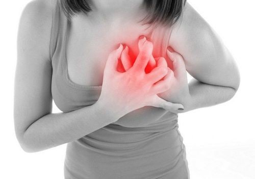
2.1 Based on clinical symptoms Symptoms of pericardial calcification are often similar to those of heart failure. They tend to occur when the patient has constrictive pericarditis. Common symptoms include:Fatigue;

Vôi hóa màng ngoài tim có triệu chứng gần giống như suy tim
2.2 Based on subclinical techniques Echocardiogram: is a non-bleeding method of functional exploration, capable of helping doctors directly observe the structures of the heart, the movement of the heart valves, the heart wall. , can determine the size of the heart chambers, the thickness of the heart wall, the pericardium, evaluate the left ventricular function,... Time Motion ultrasound can detect the thickened pericardium shown in 2 parallel lines of 2 parietal and visceral layers on the pericardium. Between these two layers, there is an ultrasonic gap of 1mm or more. From an ultrasound, the doctor can detect a thickened, calcified pericardium; X-ray: Before taking X-ray of pericardial calcification, it is necessary to aspirate fluid in the pleural space and peritoneal cavity to facilitate easy recording of X-ray images. In many cases, the pericardium is calcified - this is evidence of adhesions, thickening, and calcification. Calcification is clearly seen on lateral and longitudinal films anteriorly, above the diaphragm;
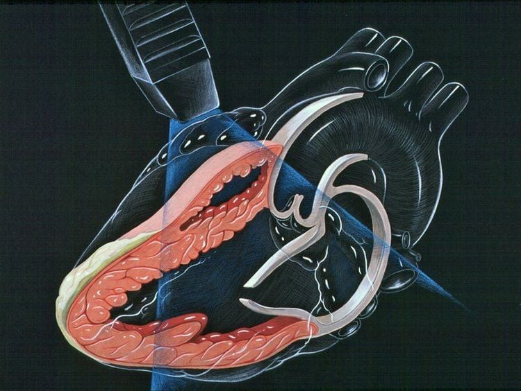
Tình trạng bệnh được chẩn đoán và đánh giá bằng các kỹ thuật cận lâm sàng
To protect cardiovascular health in general and detect early signs of cardiovascular disease, customers can sign up for Cardiovascular Screening Package - Basic Cardiovascular Examination of Vinmec International General Hospital. The examination package helps to detect cardiovascular problems at the earliest through tests and modern imaging methods. The package is for all ages, genders and is especially essential for people with risk factors for cardiovascular disease.
Please dial HOTLINE for more information or register for an appointment HERE. Download MyVinmec app to make appointments faster and to manage your bookings easily.


