This is an automatically translated article.
The article is professionally consulted by Specialist Doctor I Tran Quoc Vinh - Emergency Doctor - Department of Resuscitation - Emergency - Vinmec Nha Trang International General Hospital. Doctor Tran Quoc Vinh has more than 6 years of experience (starting in 2011) in the field of Emergency Medicine.The heart is an organ that plays a very important role in carrying blood and oxygen to nourish the whole body. The heart's blood pumping cycle is likened to "a power plant" providing 5-6 liters of blood per minute to maintain human life.
1. Structure and main function of the heart
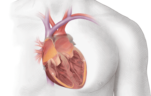
The heart is one of the most important organs in the human body. It is made up of a special type of muscle, called the myocardium. Normally, the human heart is divided into 4 parts, including:In the upper half: the left atrium and the right atrium. The common feature of these two atria is that they have thin walls, separated by the atrial septum, the right atrium receives blood from the superior and inferior vena cava to the right ventricle, and the left atrium receives blood returning from the lungs. down to the left ventricle. In the lower half: left ventricle and right ventricle. The ventricles usually have thick walls, separated by the interventricular septum, which is responsible for pumping blood into the arteries. The right ventricle pumps blood into the pulmonary artery so that the blood receives oxygen and expels carbon dioxide, the left ventricle pumps blood to the aortic arch for blood to circulate throughout the body. The heart is an essential part of the cardiovascular system. It pumps oxygen and nutrient-rich blood throughout the body to sustain life. This fist-sized heart beats continuously (opens and closes) about 100,000 times per day, pumping 5-6 liters of blood per minute, or about 2,000 gallons per day.
Trắc nghiệm: Sự thật về trái tim của bạn
Tim là 1 phần của hệ tuần hoàn, được cấu thành bởi tâm thất, tâm nhĩ, các van tim, động mạch và tĩnh mạch. Trái tim có chức năng lưu thông máu giàu oxy đưa tới khắp cơ thể. Được xem là "chìa khóa" của sự tồn tại nên việc giữ gìn một trái tim khỏe mạnh là rất quan trọng. Cùng tìm hiểu xem bạn đã thực sự hiểu về trái tim hay chưa.
Bài dịch từ: webmd.com
2. Location and shape of the heart
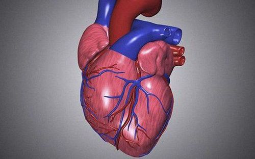
The heart is located in the middle space of the mediastinum in the thoracic cavity. It is located below the rib cage, to the left of the sternum, and in the middle of the lungs.From the outside, you can easily see that the heart is made up of heart muscle. These heart muscles contract vigorously and pump blood to the rest of the body. On the surface of the heart are the coronary arteries, which supply oxygen-rich blood to the heart muscle. In addition, the main blood vessels that enter the heart include the superior vena cava, the inferior vena cava, and the pulmonary vein. When the pulmonary artery leaves the heart, it carries low-oxygen blood to the lungs, while blood from the aorta exits and carries oxygen-rich blood to the rest of the body.
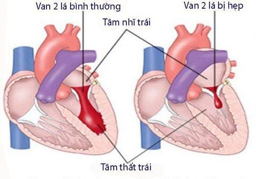
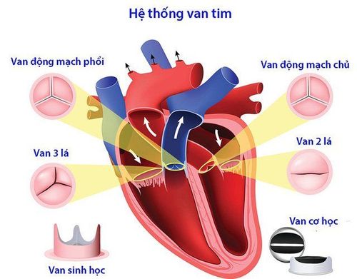
Inside, the heart is divided into 4 hollow chambers, separated by a septum on both sides. The left and right halves of the heart are divided into two chambers, the atrium in the top chamber and the ventricle in the bottom chamber. These two chambers work in conjunction with each other, contracting and relaxing to pump blood to nourish the body. As it leaves each chamber of the heart, blood must pass through the heart valves. There are usually four types of heart valves, including the mitral, tricuspid, aortic, and pulmonic valves. In which, the tricuspid valve is located between the right atrium and the right ventricle, the mitral valve is located between the left atrium and the left ventricle; The pulmonary and aortic valves are located between the ventricles and major blood vessels.
The heart valves work like a one-way valve, ensuring blood always flows in the right direction. Each valve will have a separate set of caps, also known as buttons or foils. Mitral valves usually have two leaflets, others will have three leaflets. The leaflets are attached to a ring of hard tissue called the Annulus (which maintains the proper shape of the heart valves).
3. How does the heart work?
The electrical system of the heart acts as the main source of energy that helps the ventricles and atria work alternately and relax together so that the pumping of blood through the heart occurs in the right cycle.In addition, the heartbeat will be triggered by electrical impulses traveling down a special pathway through the heart:
The electrical impulse will begin with a small bundle of specialized cells called the sinus node (SA- central node, located in the right atrium). The SA node is like a natural pacemaker, with a normal rate of 60-100 beats per minute. The electrical impulse then travels through the surrounding muscles and causes the atria to contract. At the heart of the heart, between the ventricles and atria, is a cluster of cells called the atrioventricular (AV) node. This node has the ability to slow down electrical signals before they enter the ventricles. This gives the atria time to contract before the ventricles kick in. The His-Purkinje network creates a bridge that helps the fibers send electrical impulses to the muscular walls of the ventricles, which in turn help the ventricles contract. When at rest, the heart beats about 50-99 times per minute. If you exercise, have a fever, take certain medications, or have emotional or psychological problems, your heart can beat faster than usual (more than 100 beats per minute).
4. How does the heart pump blood through the human body?
When the heart beats, some blood is pumped through a system of blood vessels, also known as the circulatory system. In it, the muscular tubes and elastic vessels will take care of the function of bringing blood to all parts of the body.As can be seen, blood plays an extremely important role in the circulatory system. Not only does it carry fresh oxygen from the lungs and other essential nutrients to the body's tissues, but at the same time, it also carries out the task of removing waste products from the tissues, including carbon dioxide. This helps maintain life and strengthens the entire body.
The human body has three main blood vessels, including:
Arteries: Starting with the aorta is considered the large artery leaving the heart. This artery carries oxygen-rich blood from the heart to all tissues in the body. They are branched many times and get smaller and smaller as they carry blood from the heart into the organs. Capillaries: These are small, thin blood vessels that connect arteries to veins. The thin-walled structure allows nutrients, oxygen, carbon dioxide and other waste products to easily pass through the body's cells. Veins: Consists of blood vessels that carry low-oxygen blood to the heart and remove waste products from the body. The closer you get to the heart, the larger the veins will become. In particular, the superior vena cava is responsible for bringing blood from the head and arms to the heart, the inferior vena cava will work and bring blood from the legs and abdomen to the heart.
5. The heart's blood pumping cycle
A circulatory system will be repeated many times, helping blood flow continuously to the heart, lungs and other parts of the body. This cycle goes like this:On the right side of the heart
Blood is brought into the heart through two large veins, the superior and inferior vena cava. It then empties the oxygen-poor blood from the body into the right atrium of the heart. When the atrium contracts, the tricuspid valve opens and blood flows from the right atrium into the right ventricle. When the ventricles are full, the tricuspid valve closes to prevent blood from flowing back into the atria during the time the ventricles contract. After the ventricles contract, the pulmonary valve moves blood out of the heart into the lungs. Here, the blood is oxygenated, expels CO2 and receives O2, then returns to the left atrium through the pulmonary veins. Left side of the heart
At this point, the pulmonary veins empty oxygen-rich blood from the lungs into the left atrium of the heart. When the atria contract, the mitral valve opens, allowing blood to flow from the left atrium into the left ventricle. After the ventricles are full, the mitral valve closes, preventing blood from flowing back into the atria while the ventricles contract. After the ventricles have contracted, the aortic valve sends blood out of the heart to all parts of the body. As blood passes through the pulmonary valve, it continues into the lungs. This process is called pulmonary circulation. Blood comes from the pulmonary valve, then travels to the pulmonary artery and small capillaries inside the lungs. Here, oxygen travels from tiny air sacs called alveoli in the lungs and through the walls of capillaries into the bloodstream. Meanwhile, the waste carbon dioxide produced through metabolism moves from the blood into the air sacs and out of the body when you exhale. After the blood has been purified and oxygenated, it returns to the left atrium through the pulmonary veins.
Vinmec International General Hospital is one of the general hospitals with the function of examination, treatment and prevention of many diseases. With a team of leading medical doctors, a system of modern equipment and technology, professional international medical examination and treatment services; A civilized, polite, safe and sterile medical examination and treatment space will bring satisfaction to customers.
Please dial HOTLINE for more information or register for an appointment HERE. Download MyVinmec app to make appointments faster and to manage your bookings easily.
Reference source: webmd.com


