This is an automatically translated article.
The article is professionally consulted by Master, Doctor Do Nguyen Thuy Doan Trang - Head of Extracorporeal Circulation Team - Cardiovascular Center - Vinmec Central Park International General Hospital. The doctor is a leading expert in Extracorporeal Circulation in cardiac surgery and cardiac resuscitation, Cardiovascular medical treatment.1. Structure of the heart
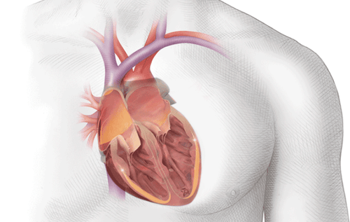
The heart is located below the ribcage, slightly to the left of the sternum and between the lungs. On the outside of the heart, the heart is made up of muscles, within the powerful muscles that contract, pumping blood to the rest of the body. On the surface of the heart, there are coronary arteries, which supply oxygen-rich blood to the heart muscle itself. The main blood vessels that enter the heart are the superior vena cava, the inferior vena cava, and the pulmonary vein. The pulmonary artery leaves the heart and carries low-oxygen blood to the lungs. The blood then exits the aorta and carries oxygen-rich blood to the rest of the body.Inside, the structure of the heart has 4 cavities, hollow, divided into two sides separated by a septum. Each side of the heart is divided into two chambers atrium and ventricle that perform the respective functions of receiving blood from the veins and pumping blood into the arteries.
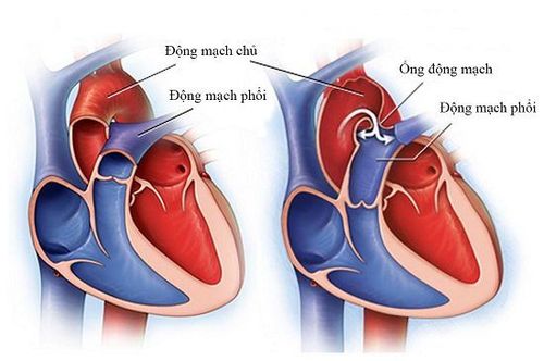
Mitral valve Tricuspid valve Aortic valve Pulmonary valve where the tricuspid and mitral valve are located between the atria and ventricle. The aortic and pulmonary valves are located between the ventricles and the main blood vessels leaving the heart.
2. How does blood pass through the heart?
The structure of the heart beats and pumps blood through a system of blood vessels – this is the circulatory system, here there are elastic vessels, constricted tubes that carry blood to all parts of the body. Besides carrying oxygen from the lungs and nutrients to body tissues, it also carries body waste products including carbon dioxide out of the tissues. This is to maintain life and promote the health of all parts of the body.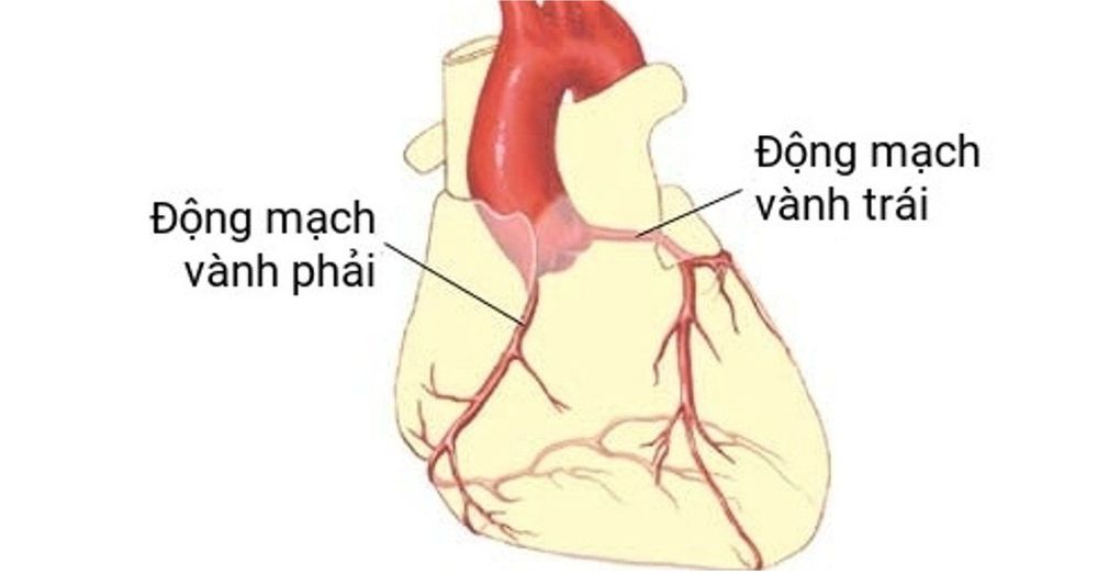
Arteries: The heart's structure begins with the aorta, which is the large artery leaving the heart. Arteries carry oxygen-rich blood from the heart to body tissues, branching many times and becoming smaller as they carry blood from the heart to specific organs in the body. Capillaries: Small, thin blood vessels that connect arteries and veins together, they allow oxygen, nutrients, carbon dioxide, and other waste products to pass through and from the cells of the organ. Veins: As the blood vessels that bring blood back to the heart, they have a lower oxygen content and are rich in waste products that will be eliminated or eliminated from the body.
3. How does blood flow through the heart?
The right and left sides of the heart work together, this process repeats, causing blood to flow continuously to the heart, lungs and organs.On the right side of the heart:
Blood enters the heart through two large veins, the inferior and superior vena cava, which carry low-oxygenated blood from other parts of the body into the right atrium of the heart. When the atria contract, blood flows from the right atrium into the right ventricle of the heart through the open tricuspid valve.
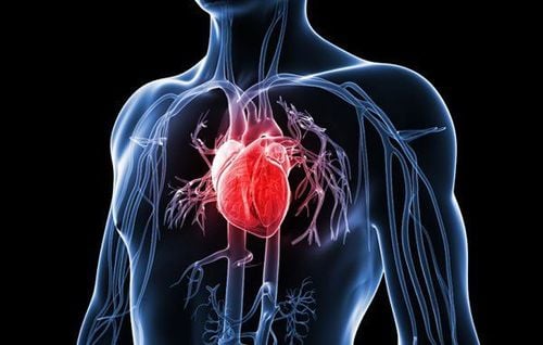
The pulmonary veins transport blood with oxygen-rich blood from the lungs into the left atrium of the heart When the atrium contracts, blood flows from the left atrium of the heart into the left ventricle through the mitral valve open When the heart When the ventricles are full, the mitral valve closes to prevent blood from flowing back into the atria when the ventricles contract. When the ventricles contract, blood exits the left ventricle through the aortic valve, into the aorta, and into the body. To protect heart health in general and detect early signs of myocardial infarction and stroke, customers can sign up for the Cardiovascular Screening Package - Basic Cardiovascular Examination of Vinmec International General Hospital. The examination package helps to detect cardiovascular problems at the earliest through tests and modern imaging methods. The package is for all ages, genders and is especially essential for people with risk factors for cardiovascular disease.
Please dial HOTLINE for more information or register for an appointment HERE. Download MyVinmec app to make appointments faster and to manage your bookings easily.


