This is an automatically translated article.
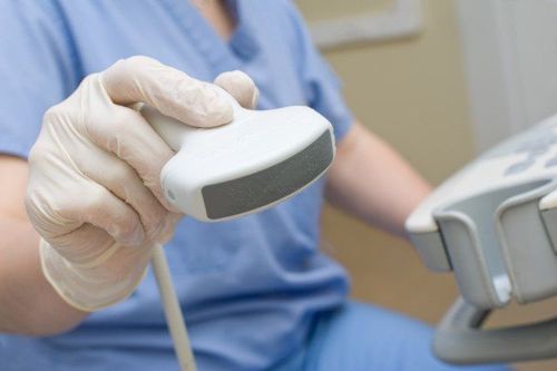
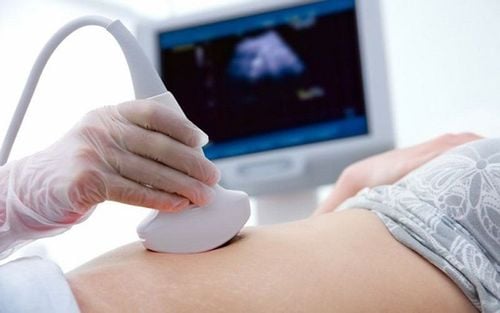
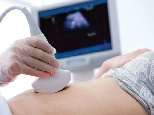
The article is professionally consulted by Master, Doctor Nguyen Viet Thu - Doctor of Radiology and Nuclear Medicine - Department of Diagnostic Imaging and Nuclear Medicine - Vinmec Times City International General Hospital.Over the past 40 years, ultrasound has become an important diagnostic modality. The use of ultrasound in medicine began during and shortly after World War II. This article will show how ultrasound technology came to be and some of its notable contributions to the field of medicine. ultrasound technique.
1. Ultrasound technique
Ultrasound technology in medicine is constantly evolving and plays an important role in the diagnosis and treatment of diseases. But few people know how the history of ultrasound technology was born and developed? To find out, let's look back at the more than 225-year history of ultrasound.In 1942, neurologist Karl Dussik used ultrasound to find the location of tumors in his patient's brain and he is considered the first person to use ultrasound in medicine. However, the applications of ultrasound were actually discovered and put into practice long before that.
In 1794 physiologist, professor and priest named Lazzaro Spallanzani in a study of bats discovered that this animal has the ability to adjust flight direction to avoid obstacles by sound waves of high frequency. height they emit, not visually. The reflected waves return to help them shape the distance and size of the obstacle to give the right flight direction. This is considered the initial foundation to put ultrasound into practice.
In 1877, Pierre and Jacques Curie invented the piezoelectric effect, which was later used to create ultrasonic transducers.

Đầu dò siêu âm được Pierre và Jacques Curie phát minh ra từ hiệu ứng điện áp
In the 1920s - 1940s, ultrasound was used as a form of physical therapy to relieve pain and restore muscle and joint mobility for European football players.
In 1948 George D. Ludwig, an internist at the Naval Medical Research Institute, invented an ultrasound device that he named the A-mode to detect gallstones.
1945 - 1951 Douglas Howry and Joseph Holmes from the University of Colorado developed the B-mode ultrasound device. John Reid and John Wild improved and used this device to diagnose breast cancer.
In 1953, doctors Inge Edler and Hellmuth Hertz successfully performed the first echocardiogram, marking a turning point for the application of ultrasound in medicine later.
In 1966 Don Baker, Dennis Watkins and John Reid invented the Doppler ultrasound machine to help display images of blood flowing in the blood vessels. Later in the 1970s, the continuous Doppler ultrasound technique evolved into continuous wave doppler, spectral doppler, and color doppler ultrasound.
In the 1980s, Kazunori Baba of the University of Tokyo developed 3D ultrasound technology and first captured 3D ultrasound images of the fetus in 1986.
In 1989 Professor Daniel Lichtenstein started combining Ultrasound in general and lung ultrasound in particular in diagnosis and treatment.
Since then, ultrasound techniques have been continuously improved, developed and applied in many different fields in medicine. Appearance of color ultrasound, 4-dimensional ultrasound (a type of video describing the shape of organs, organ systems and their movements in the body),... Ultrasound is also applied to many procedures such as childbirth device or endoscopy.
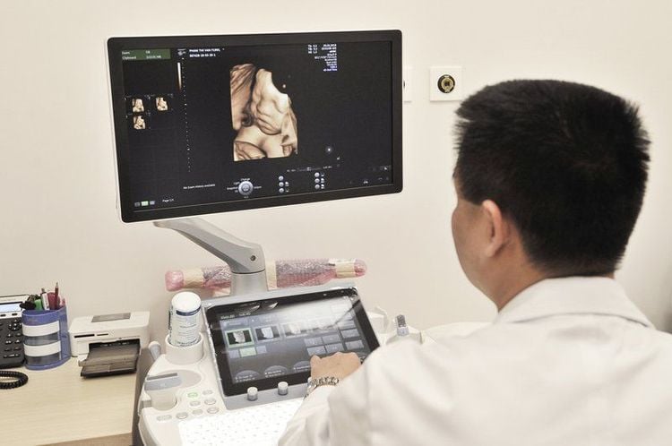
Siêu âm 4D ngày càng được ứng dụng rộng rãi nhất là trong siêu âm thai nhi
2. Application of ultrasound in the medical industry
In medicine, ultrasound uses sound waves with frequencies from 3 to 10 MHz. The most important parameters in ultrasound are: Wavelength, frequency, velocity, intensity are linked together by the formula:v = fl
Where:
v is the speed of the sound wave
f is the frequency negative wave number in Hz
l is wavelength
In diagnosis and treatment, short wavelength sound waves are commonly used. Human tissues and organs are not homogeneous, so the reception, absorption and reflection of sound waves are also different.
Acoustic reflection is highly dependent on the resistance of individual tissues or organs. For abdominal ultrasound such as liver, kidney, pancreas, the wave frequency is used around 3MHz, while for some other parts such as neck, breast, this number needs to reach 5MHz or even 7MHz to get a clear image. and exactly.
The application of ultrasound in medicine is huge. There are many ultrasound modes applied in medicine so far, typically the following 4 modes:
● Mode A: Ultrasound mode A is the simplest type of ultrasound. The transducer will cut a slice through the body to show images of tissues, organs, and internal organs. In addition, therapeutic ultrasound is also a form of this regimen.
B mode: Show 2D ultrasound images on the screen
M mode: Can display the movement of tissues, organs, internal organs on the mechanism using B mode ultrasound changes change the probe scan time and speed.
● Doppler ultrasound: Used to measure and display blood flow in the vessel. Doppler ultrasound plays an important role in the diagnosis of several cardiovascular diseases.

Siêu âm Doppler là một chế độ siêu âm được áp dụng trong y học từ trước đến nay
Ultrasound since its appearance has continuously developed and widely applied in the fields of medicine. Ultrasound techniques and equipment are increasingly improved, from color ultrasound, 3D ultrasound, 4D ultrasound, Doppler ultrasound,... This helps the applications of ultrasound in the medical industry more and more. richer. Not only can ultrasound simply capture images of the body's tissues, but now it can also represent those images in the most realistic way, assisting both in the rehabilitation process and in procedures performed in the hospital. surgery.
Currently, there are many medical facilities that apply ultrasound technology in the early diagnosis and treatment of diseases. Vinmec International General Hospital system has been using the most modern generations of color ultrasound machines today in examining and treating patients. One of them is GE Healthcare's Logiq E9 ultrasound machine with full options, HD resolution probes for clear images, accurate assessment of lesions. In addition, a team of experienced doctors and nurses will greatly assist in the diagnosis and early detection of abnormal signs of the body in order to provide timely treatment.
Ths.Bs Nguyen Viet Thu has more than 20 years of experience working in the field of diagnostic imaging, former Secretary of the Department of Radiology in Hanoi, directing the lower level in the field of Diagnostic Imaging. Currently, the doctor is working at the Department of Diagnostic Imaging and Nuclear Medicine - Vinmec Times City International General Hospital.
Customers can directly go to Vinmec Health system nationwide to visit or contact the hotline here for support.
Source: ultrasoundschoolsinfo.com
SEE MORE
Evaluation of pulmonary artery pressure by echocardiography Doppler echocardiography Application of echocardiography What you need to know about echocardiography
