This is an automatically translated article.
Congenital duodenal obstruction is a common intestinal malformation, occurring in 1 in 5,000 - 10,000 live births. It causes intermittent or intermittent vomiting within the first 24 to 38 hours postpartum, usually after the first milk feed. The definitive intervention method to correct congenital duodenal obstruction in children is surgery.1. Causes of congenital duodenal obstruction
Congenital duodenal obstruction is an obstructed duodenal condition that develops before birth. The underlying cause is unknown, but genetics may play a role. No maternal predisposing risk factors were found.
The incidence of congenital duodenal obstruction is about 1 in every 5,000 to 10,000 live births, more common in boys than girls. Duodenal obstruction may be a separate condition, or occur in conjunction with other related birth defects. Although 25-40% of patients with congenital duodenal obstruction have Down syndrome, this condition is not an independent risk factor for the development of duodenal obstruction.
Duodenal obstruction is associated with VACTERL, annular pancreas, and other intestinal obstructions such as jejunal obstruction, rectal obstruction.

Tắc tá tràng bẩm sinh có thể là một dị tật bẩm sinh
2. Prognosis of children with congenital duodenal disease
"How dangerous is congenital duodenal obstruction?" According to the US National Institutes of Health, when congenital duodenal obstruction is diagnosed and treated promptly, the prognosis is very good, most children grow up with normal lives.Despite improvements in early mortality, up to 22% of children with duodenal obstruction may develop late complications after surgery. Late complications include blind loop syndrome, giant duodenum, gastroduodenal reflux gastritis, peptic ulcer, esophagitis and gastroesophageal reflux disease, pancreatitis, and cholecystitis. .
Studies show that the mortality rate is about 3%. Most deaths and complications occurring in pediatric duodenal obstruction are attributed to the presence of other associated malformations (usually complex cardiac malformations).
3. Prenatal symptoms of duodenal obstruction.
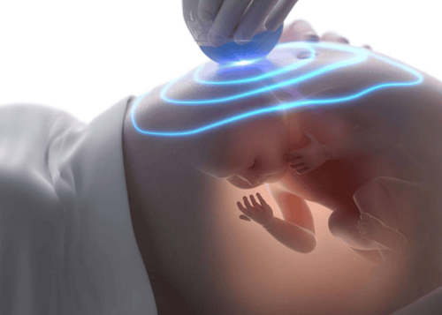
Congenital duodenal obstruction can be diagnosed by ultrasound during the third trimester of pregnancy. Perform screening for duodenal obstruction if the baby is at risk for Down syndrome or the mother has polyhydramnios. Note that normal ultrasonography does not rule out congenital duodenal obstruction.Signs on prenatal ultrasound help diagnose the disease:
Polyhydramnios: Congenital duodenal obstruction often leads to polyhydramnios, if polyhydramnios is present, the sonographer suggests duodenal obstruction. In a normal pregnancy, the fetus will swallow amniotic fluid, but when the duodenum is blocked, it will be difficult for the fetus to absorb, leading to accumulation of amniotic fluid. Polyhydramnios has the potential to increase the risk of complications during pregnancy, such as premature birth. Double shadow sign: This is the classic sign of duodenal obstruction on sonography, characterized by two shadows corresponding to the stomach and duodenum filled with fluid. This sign indicates an accumulation of fluid due to a blockage. More than 50% of babies with duodenal obstruction have other birth defects, so they will have fetal echocardiography and amniocentesis to determine if the baby has Down syndrome or other birth defects.
Prenatal diagnosis helps consult with a pediatric surgeon about treatment as well as provides parents with a discussion of a postpartum care and management plan.

Tắc tá tràng bẩm sinh sẽ được điều trị tốt nhờ chẩn đoán trước khi sinh
4. Postpartum symptoms of congenital duodenal obstruction.
If congenital duodenal obstruction is not diagnosed during pregnancy, it usually manifests clinically in the first 24 to 38 hours after birth.Newborns may vomit after the first feed, and the vomiting gets worse over time. Repeated vomiting, or green-colored vomit, is a sign that the bowel obstruction is located farther than the tube of Vater. The child may also be bloated and not have a bowel movement.
Abdominal radiograph: When duodenal obstruction is suspected, an upright abdominal radiograph is the first imaging modality used after birth. The characteristic sign of duodenal obstruction is the double silhouette of an air-filled stomach and the first part of the duodenum. Cardiac and renal echocardiography: Performed if the infant has not been evaluated for cardiac and renal abnormalities before birth, helps identify life-threatening abnormalities before surgery for duodenal obstruction. Contrast-enhanced gastroscopy is sometimes used to evaluate the esophagus, stomach, and duodenum. The amount of barytium deposited indicates interference. The primary purpose of imaging is to differentiate between duodenal obstruction and transverse colonic volvulus; an important distinction, because transverse colonic volvulus requires urgent surgery. Pediatric CT plays a very limited role, if any, in the diagnosis and evaluation of duodenal obstruction. However, CT reconstruction allows further assessment of bowel distribution in some difficult cases.
5. Treatment of congenital duodenal obstruction
Duodenal obstruction is treated after the baby is born, but there are several prenatal interventions that reduce the risk of birth complications such as amniocentesis to drain fluid in the case of polyhydramnios. Close monitoring is necessary so that the fetus and mother can be monitored to detect emergency problems early.Babies diagnosed with congenital duodenal obstruction can be delivered normally. The general goal is to have a normal delivery as close to the due date as possible. The birth process can take place normally, but requires intensive medical interventions after the baby is born, the baby will be taken to the intensive care room.
Babies born with a diagnosis of congenital duodenal obstruction are fed parenteral nutrition. In addition, a nasogastric tube is inserted into the child's stomach, helping to clear trapped gas and fluid in the stomach. Your baby will not be bottle-fed or breast-fed until the surgery is done.
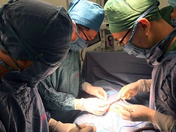
Surgery is the definitive treatment for duodenal obstruction in infants, which can be performed either open or laparoscopically. Surgery is required, but emergency surgery is usually not necessary, except when the child has a metabolic disorder or bowel obstruction. Allow a 24- to 48-hour delay to facilitate further assessment and fluid resuscitation.

Phẫu thuật trị dứt điểm tắc tá tràng bẩm sinh
Please dial HOTLINE for more information or register for an appointment HERE. Download MyVinmec app to make appointments faster and to manage your bookings easily.


