This is an automatically translated article.
The article is written by Master, Doctor Mai Vien Phuong - Gastroenterologist - Department of Medical Examination & Internal Medicine - Vinmec Central Park International General Hospital.
Histopathology is the gold standard for diagnosing intrahepatic hemangiomas, because imaging and clinical features are difficult. Notably, histological analysis of liver biopsies also showed that the misdiagnosis of intrahepatic hemangiomas was in about 15% of cases.
1. Overview
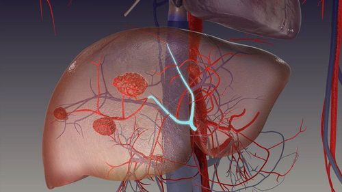
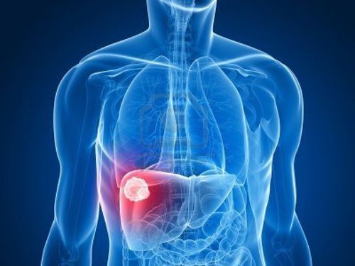
First reported in 1976, intrahepatic hemangioma (HAL) is a rare liver mesenchymal tumor that mainly occurs in middle-aged women. Diagnosis of a liver tumor is often incidental on abdominal ultrasonography during physical examination in the absence of specific symptoms.
Almost 10% of intrahepatic hemangiomas are associated with tuberous sclerosis complex. Liver hemangiomas contain varying proportions of blood vessels, smooth muscle cells, and adipose tissue, making radiographic diagnosis difficult. The cells expressed positivity for HMB-45 and actin, thus these tumors were integrated into the group of perivascular epithelial cell tumors.
Liver hemangioma is a rare, but not exceptional, liver mesenchymal tumor. Hemangiomas in the liver contain different proportions of blood vessels, smooth muscle cells, and adipose tissue, making its imaging-based diagnosis difficult. In most cases, after a benign clinical course, the tumor is potentially malignant, complicating management, which is still less codified.
2. Clinical features of liver hemangiomas
Intrahepatic hemangioma is a common tumor in the liver, without cirrhosis, and mainly affects middle-aged women. A retrospective analysis of the literature performed up to 2016 identified 292 patients with one or more hepatic hemangiomas, most of whom (almost 74%) were women, with a mean age of 24 years and older. to 53.
Intrahepatic hemangiomas were mainly located in the right liver (60% of cases), isolated in 84% of cases, the median size ranged from 2 to 12.7 cm. This type of tumor is often discovered incidentally during physical examination (42% to 72% of cases) because most subjects are asymptomatic. Symptoms of liver hemangioma may include:
Abdominal pain or discomfort. Abdominal bloating, weight loss. Rarely, an abdominal mass is found on palpation. A few cases of spontaneous rupture of intrahepatic hemangiomas have been reported. Tumor size (≥ 4 cm) and pregnancy are two conditions recognized in favor of Rupture of Renal Angiomyolipoma, but the small number of reported hepatic hemangioma ruptures preclude the identification of predisposing factors. rupture in the liver.

3. Histology is the gold standard for diagnosing hemangiomas in the liver
Histology is the gold standard for the diagnosis of hemangiomas in the liver, because imaging is difficult. Notably, histological analysis of liver biopsies also showed that intrahepatic hemangioma was misdiagnosed in about 15% of cases in a recent multicenter study.
The World Health Organization defines a perivascular epithelial cell tumor as “a mesenchymal tumor containing distinctive perivascular epithelial cells”. Angiomyolipoma belongs to the group of PEComa, consisting of adipose tissue, smooth muscle, and vessels that can become dystrophy. Histological and immunohistochemical characteristics classify hepatic hemangiomas in the group of perivascular ependymoma, a group of tumors with different histological characteristics, but a common immunohistochemical marker, specifically may be co-expression of melanocyte and muscle markers.
The PEComa family includes Angiomyolipomas, clear-cell "sugar" tumors of the lung, lymphangiomas (LAMs) and a variety of visceral, intra-abdominal and soft-tissue/skeletal abnormalities, described under the term "PEComa". clear myeloid tumor of the ligament of falciform/Ligamentum teres". There is a strong association between Angiomyolipoma and tuberous sclerosis complex, and between LAM and tuberous sclerosis complex, although this association is less marked for other members of the PEComa family.
4. Types of cells in liver hemangiomas
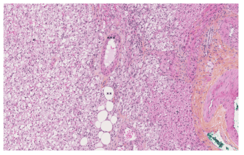
Perivascular epithelial cells are characterized by their perivascular location, often with a radial arrangement of cells around the vessel. Normally, these cells are predominantly epithelial as they are only perivascular, whereas smooth muscle-like rhabdoid cells are seen further distal to the vessel. In the liver, the aspect is usually only the epithelium without the rhabdoid cells.
Fat cells are usually found far from blood vessels. Wide differences are seen in the relative proportions of epithelial, rhabdoid, and lipid puffy cells. Depending on the relative proportions of these different tissues, on the one hand, there is a common Angiomyolipoma with a predominance of cells, fat vessels, or muscle, and on the other hand, an epithelial Angiomyolipoma containing at least 10% of epithelial cells. Mixed angiomyolipoma and myofibroma were the most common (36% and 42% of the 151 cases of hepatic hemangioma for which informative data were available, respectively).
Another subtype of intrahepatic hemangioma, inflammatory hepatic hemangioma, has also been recognized, although only 14 cases have been reported in the English literature. Overall, this tumor is characterized by an excessive inflammatory infiltrate (50% of the tumor area), the main inflammatory cell types being lymphocytes (100%), plasma cells (93%) and histiocytosis. (71%). Macroscopically, intrahepatic hemangiomas are well-defined, uncapsulated, smooth and brown in color; however, the existence of hemorrhage or necrosis within the tumor may change its shape. On microscopic examination, there are usually clear, small, central, round or oval eosinophils, with a small nucleus. "Atypical" angiomyolipomas exhibit cytologic loss, multinucleated nuclei, focal necrosis, and increased mitotic counts.
5. Differential diagnosis of liver hemangiomas on histopathology
There can be many differential diagnoses of perivascular ependymoma, depending on the location and predominant tissue that constitutes the tumor. With uniform expression of melanoma markers, periangiomas can be confused with common melanoma and clear cell sarcoma, but this type of malignancy often presents Strong expression of S100 protein and no smooth muscle actin staining.
Because of the favorable intra-abdominal location, presence of epithelial cells, rhabdoid cells, and occasional positive KIT staining (CD117) in hepatic hemangiomas, the diagnosis of gastrointestinal stromal tumor is sometimes Be discussed. Depending on the size of the contingent of epithelial, rhabdoid or adipocytes, Angiomyolipoma can also be confused with carcinoma, leiomyoma, or lipoma
In short, angiomas Blood in the liver is a rare but not exceptional tumor and usually has a benign course. However, this tumor may present a more malignant potential when it recurs or metastasizes. Therefore, in some cases, the liver hemangioma is large and causes clinical manifestations such as right upper quadrant pain, the tumor grows and causes significant damage to the healthy liver parenchyma. hepatic hemangioma, radiotherapy,...
Although there are no strong histological or radiological features to predict the natural course of this tumor type. Diagnosis by radiographic imaging is often difficult because of the variable incidence of liver hemangioma-containing tissues. Therefore, histological analysis of the tumor and multimodal consultation, whenever possible in a specialist center, are essential for optimal care of these patients.

6. Relationship between tuberous sclerosis complex (TSC) and liver hemangioma
The relationship between tuberous sclerosis complex (TSC) and renal angiomyolipoma, first described in 1911, was observed in 50% of cases, while the association between tuberous sclerosis complex and hepatic hemangioma was only observed in 50% of cases. is observed in 5-15% of cases.
Tuberous sclerosis complex is an autosomal recessive disorder with a neonatal incidence of 1:6000, although sporadic cases of de novo mutations are the most frequent presentation in the absence of a family history. family. Tuberous sclerosis complex results from mutations in tuberous sclerosis complex 1 or tuberous sclerosis complex 2, which code for hamartin and tuberin, respectively. These proteins are important regulators of cell growth and proliferation, likely through their upstream modulator, the mammalian rapamycin (mTOR) target. Loss of function or dysfunction of either protein leads to the development of hamartomas in many organ systems, including the brain, kidneys, heart, and liver.
In patients with tuberous sclerosis complex, hepatic hemangiomas are often associated with renal angiomyolipoma. A recent retrospective study showed that, among 25 hepatic hemangiomas, 88% also had renal angiomyolipoma, and patients with tuberous sclerosis complex 2 had a higher frequency of hepatic hemangiomas than patients with complex sclerosis. tuber 1 (18% vs 5%; P = 0.037). In contrast to previous reports, female predominance was observed in patients with intrahepatic hemangiomas but without tuberous sclerosis complex.
7. Prognosis of hepatic hemangioma
The scarcity of perivascular ependymoma precludes the identification of robust criteria to differentiate benign Angiomyolipoma from other potentially more malignant neoplasms. The first description of a potentially “malignant” intrahepatic hemangioma was recent, and although the authors did not explicitly indicate the malignant nature of this tumor, the reported features (ie. are large size, loss of cytology and presence of necrosis), tumor, and patient-related mortality are strong arguments in favor of the "malignant" case.
From a series of 24 periangiomas of the soft tissue and gynecologic tract (excluding Angiomyolipoma) with a mean follow-up of 30 months (range: 10-84), Folpe et al observed found 3 local recurrences and 5 distant metastases (8/24, 33% cases), 2 deaths (8%), 4 patients (17%) alive with metastatic local disease or unresectable, 18 patients (75%) were alive with no evidence of disease.
A meta-analysis of these 24 cases, plus 45 other reported cases in the literature, with adequate follow-up information available identified risks associated with increased recurrence or metastasis , which are:
Tumor size larger than 5 cm Infiltrative growth pattern High nuclear degree Necrosis and mitotic activity > 1/50 of high energy field. Thus, these authors developed a provisional classification of perivascular ependymoma with increasing malignancy. Regarding intrahepatic hemangiomas, it is mainly the epithelial type that poses the risk of malignancy. In a review published in 2017, the mortality rate associated with liver hemangiomas was 0.8%.
Classification of perivascular epithelial cell tumors according to their malignancy:
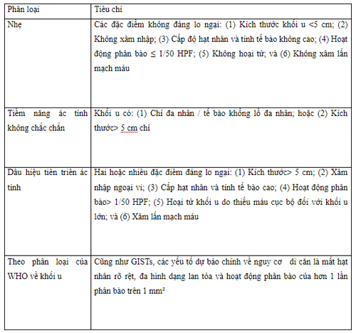
GIST: Gastrointestinal stromal tumor; HPF: High Power Microfield.
Treatment of liver hemangiomas is not usually necessary. Most diseases only need periodic monitoring is enough. However, in some cases, the liver hemangioma is large in size and causes clinical manifestations such as right upper quadrant pain, the tumor grows and causes significant damage to the healthy liver parenchyma. Liver hemangioma, radiotherapy,...
Please dial HOTLINE for more information or register for an appointment HERE. Download MyVinmec app to make appointments faster and to manage your bookings easily.
References: Calame P, Tyrode G, Weil Verhoeven D, Félix S, Klompenhouwer AJ, Di Martino V, Delabrousse E, Thévenot T. Clinical characteristics and outcomes of patients with hepatic angiomyolipoma: A literature review. World J Gastroenterol 2021; 27(19): 2299-2311 [DOI: 10.3748/wjg.v27.i19.2299]
Zhou YM , Li B, Xu F, Wang B, Li DQ, Zhang XF, Liu P, Yang JM. Clinical features of liver hemangiomas. Hepatobiliary Patients Pancreat Dis Int . 2008; 7: 284-287. [ PubMed ] [Citation in this article: 3 ]
Occhionorelli S , Dellachiesa L, Stano R, Cappellari L, Tartarini D, Severi S, Palini GM, Pansini GC, Vasquez G. Spontaneous rupture of hepatic epithelial angiomyolipoma: injury control surgery. A case report. G Chir . Year 2013; 34 : 320-322. [ PubMed ] [Quote in this article: 3 ]


