This is an automatically translated article.
The article is professionally consulted by Master, Doctor Phan Ngoc Toan - Emergency Medicine Doctor - Emergency Department - Vinmec Danang International Hospital. The doctor has a lot of experience in the treatment of Resuscitation - Emergency.1. Overview
Angioplasty site complications were defined as the presence of a hematoma at the procedural site (perivascular tissue) with or without a pseudoaneurysm, prior to hospital discharge and classified as 1 of 4 types:Mild: no treatment is needed Moderate but requires a blood transfusion Moderate but requires Thrombin injection Severe: requires surgical treatment There are many situations that lead to these complications, depending on the nature of the procedure needed. surgery – surgery (emergency, emergency, delay ..). The most common, however, is the situation involving cardiovascular interventions (pictured below).
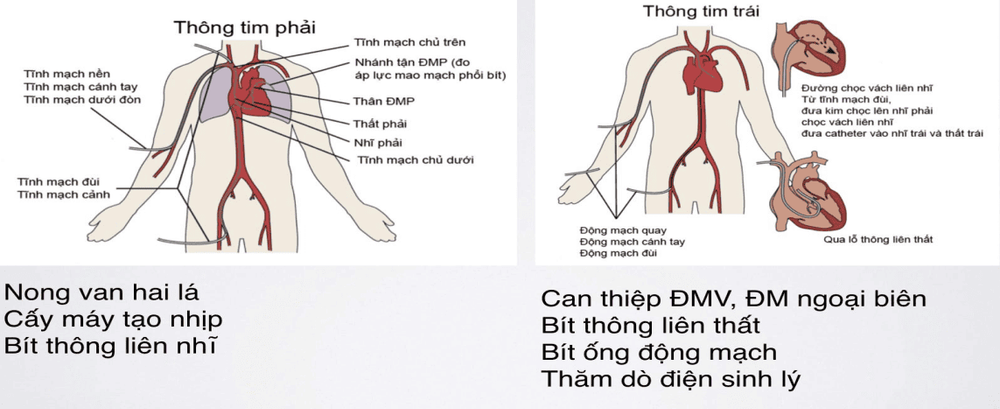
Antiplatelet drugs, anticoagulants, thrombolytics. Female Elderly, obese Peripheral artery calcification, twisting... Procedure: large sheath diameter.
2. Common Complications
Bleeding: Hematoma around the puncture site, hematoma into the following compartments: retroperitoneum, femoral cavity, forearm muscular space Pseudoaneurysm Arterial venous catheterization Thromboembolism Infection.3. Symptoms and treatment
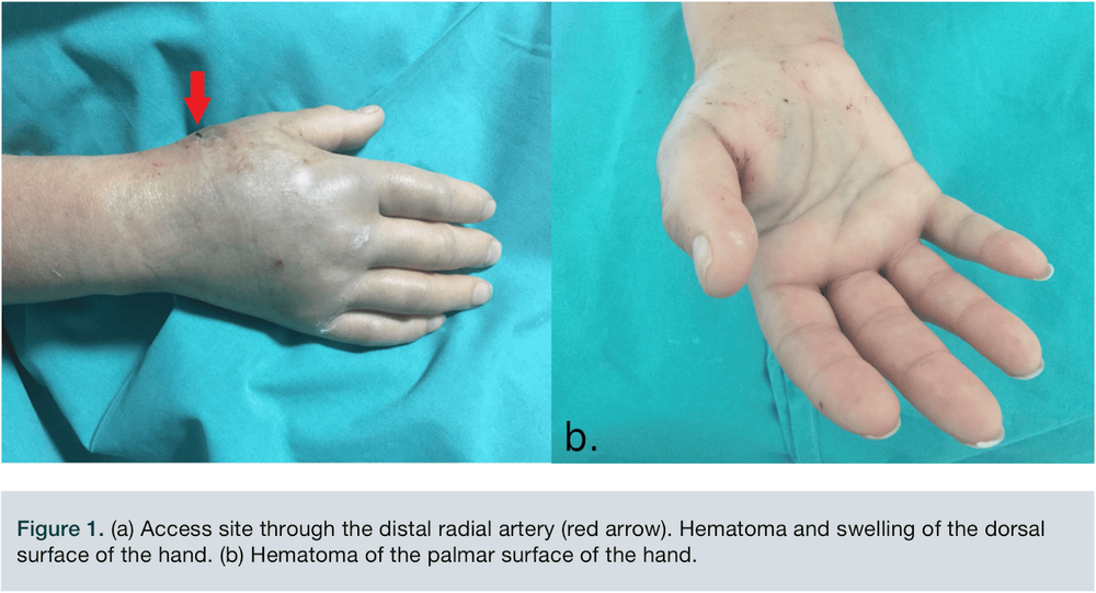
3.1. Peripheral hematoma
Painful swelling at the puncture siteSqueeze over the hematoma 1-2 cm and monitor clinically. Severe signs: Size > 4 cm, uncontrolled bleeding, tachycardia, low blood pressure 🡪
Intensive resuscitation treatment.
3.2. Retroperitoneal hematoma
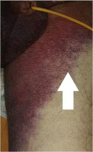
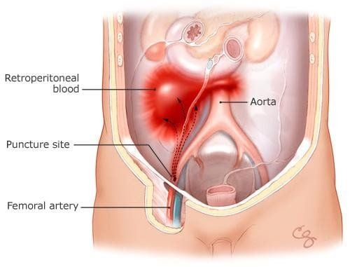
Blood flow into the retroperitoneal space Usually due to high puncture site blood flows from the back of the iliac artery gradually seeping into the retroperitoneal space Rate: 0.1-0.2% Clinical:Signs of blood loss : pale, cold Abdominal pain, low back pain Heart arrhythmia, low blood pressure, little response to fluid replacement. Cullen's sign and Gray Turner Hb decrease... Diagnosis: Abdominal ultrasound, abdominal CT with contrast can help detect the hematoma and the site of drug drainage
Management:
Pressure over 1-2 cm Transfusion, blood transfusion, neutralization of anticoagulants. The patient was hemodynamically stable, with no signs of further blood loss, and treated conservatively. Intervention: Hemodynamic instability, blood transfusion > 4 units in 24 hours Coil occludes bleeding site, Cover Stent Surgery: When other methods fail, hematoma compresses intra-abdominal organs
3.3. Ripping aneurysm:
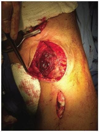
Injury to all 3 layers of vessels causes blood to flow from the lumen to an extravascular bagFrequency: 0.005-2%
Clinical:
Painful swelling at the puncture site Examination shows a heart-beating mass Management:
Squeeze over 1-2 cm for >10 minutes (average 30 minutes): successful 75-98 % Injection of coagulation drug into the site of pseudoaneurysm. Coil, Cover Stent Surgery: Other methods fail, when pseudoaneurysm infection, rapid growth, skin necrosis
Please dial HOTLINE for more information or register for an appointment HERE. Download MyVinmec app to make appointments faster and to manage your bookings easily.


