This is an automatically translated article.
The article was professionally consulted by Doctor Trinh Le Hong Minh - Radiologist - Radiology Department - Vinmec Central Park International General Hospital.Breast cancer is the most common malignancy in women. To screen and prevent cancer, it is necessary to have advanced measures to early identify cells that are at risk of developing into disease. Breast ultrasound is an imaging technique that plays an important role in detecting abnormalities in the mammary glands, especially in breast cancer screening.
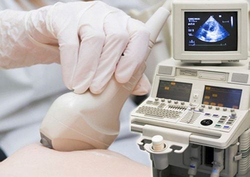
1. What is a breast ultrasound?
The mammary gland is composed of 10-15 lobules arranged in a circle, which plays the role of milk production. There are also milk ducts 0.5mm in diameter and surrounding epithelial muscle cells that help shape the breast.Breast ultrasound (also called breast ultrasound) is an imaging method widely used in medicine today. Breast ultrasound is based on the use of ultrasound waves to capture images of the internal structure of the mammary gland and body, thereby diagnosing abnormalities and diseases in the mammary gland.
Advantages of breast ultrasound:
Simple, low cost ultrasound procedure. Fast results, high accuracy of results. Noninvasive. The procedure is safe, with no pain or discomfort. There are no side effects to the patient's health. Applicable to all ages, all genders.
2. The role of breast ultrasound
In general, breast ultrasound has the following functions:Detecting abnormalities and abnormalities in the mammary glands. Distinguish between fluid-filled cysts and solid tumors in the breast. Identify small lesions (<5mm) that are normally difficult to notice. Screening for malignant breast tumors, breast cancer. Support needle guidance in invasive procedures such as cyst aspiration, biopsy, guide core biopsy...

Siêu âm vú có thể giúp phát hiện bất thường, dị thường tuyến vú
3. When to have a breast ultrasound?
Experts recommend that there are a number of groups of subjects who should pay attention and perform periodic breast screening:Women aged 35 and older should actively check and perform regular breast ultrasound every 3-6 months to early detection of the risk of breast disease. Especially those with a family history of breast cancer. Women with early menarche (under 12 years old) or late menopause (after age 55), regularly taking oral contraceptives or estrogen replacement... are at high risk of breast cancer and need regular screening. Those who do not have children or give birth to their first child after the age of 30, often eat greasy foods, drink a lot of alcohol, tobacco, and are exposed to radiation. In addition, you should also go for a breast ultrasound if you see abnormal signs that warn of cancer such as:
Unusual chest pain. Felt a lump in the chest. Swollen lymph nodes under armpit. Inflammation of the skin around the chest. The skin around the tip of the nipple changes abnormally. Nipple inward

Hãy đi siêu âm kiểm tra vú nếu bạn cảm thấy đau tức vùng ngực bất thường
4. Breast ultrasound procedure
The ultrasound technique of the breast will be scanned on the entire mammary gland, combining the two-sided comparisons, surveying the area of the lesion and surrounding tissues. In general, the breast ultrasound procedure will be conducted in turn according to the following steps:Step 1: The patient lies supine on the examination bed, arms raised (creating a 45-degree angle between the arm and forearm) for the specialist to perform the ultrasound. The sound can easily survey the inner part of the mammary gland, behind the areola, then lie sideways to examine the outside of the mammary gland.
Step 2: The doctor applies a layer of gel to the patient's chest area to help the detector make sure contact with the mammary gland, preventing air in between the detector and the human skin.
Step 3: The doctor moves the transducer along the horizontal, vertical planes from the center to the periphery or in a swirl from the nipple, areola to the outside of the mammary gland. The transducer emits high-frequency ultrasound waves that capture images of the mammary gland and display it on a monitor.
Step 4: For areas with suspected lesions, the doctor rotates the probe 90 degrees to determine on both planes of the mammary gland. Combining the technique of pressing - releasing the probe on the lesion causes morphological changes to distinguish cysts or dense fibroadenomas.
Step 5: Assess the extent of the damage, click save the image, mark the position and distance from the nipple, measure the size and number of tumors or lesions...
Step 6: The doctor wipes the substance Gel is applied initially and at the end of breast ultrasound. The ultrasound takes about 30 minutes and does not cause any pain or affect the patient.
Kỹ thuật siêu âm vú được sẽ được quét trên toàn bộ tuyến vú, kết hợp so sánh 2 bên bình diện, khảo sát khu trú vùng tổn thương và các mô xung quanh
The ultrasound process is carefully performed by the sonographers, experienced imaging specialists, with high expertise with the current ultrasound machine system. Modern, advanced technology provides detailed, clear images of mammary gland structure. Early detection of disease and signs of disease will greatly help in the treatment of breast disease later on.
Please dial HOTLINE for more information or register for an appointment HERE. Download MyVinmec app to make appointments faster and to manage your bookings easily.