This is an automatically translated article.
The article was professionally consulted with Master, Doctor Bui Ngoc Phuong Hoa - Neurologist - Department of Medical Examination & Internal Medicine - Vinmec Danang International General Hospital.Through the examination of focal neurological signs helps to identify the manifestations of dysfunction in regions of the central nervous system. From there, an accurate diagnosis can be made to provide an appropriate treatment plan.
1. Focal neurological signs
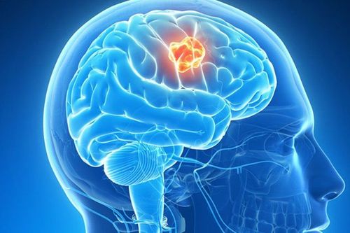
The focal nerve is the area where the nerves are gathered to form the central nervous system, including areas such as: frontal lobes, parietal lobes, temporal lobes, occipital lobes, cerebellum, and brainstem (peduncle) brain), spinal cord.Focal neurologic signs are cognitive and behavioral signs caused by focal lesions in an area of the central nervous system.
Focal neurological signs are grouped as follows:
1.1 Signs of the frontal lobes
Frontal lobe signs are often motor-related, including many characteristic defects, depending on the location of the frontal lobe affected:Unsteady walking. Stiffness, resistance to passive movement in the extremities (hypertonia). Paralysis of a limb or hemiplegia. Paralysis of eye movements. The patient has lost the ability to express in words. Seizures may be localized or diffuse to adjacent areas. Major epileptic seizures. Personality changes such as depression, ill-timed laughter, unexplained tantrums; lost initiative and interest, emotionless, speechless and motionless, stagnant. The "frontal lobe detachment" sign, for example, reappears primitive reflexes such as the nasal reflex, the grasp reflex, and the hand-chin reflex. Loss of smell on one side (anosmia).
1.2 Signs of lobes
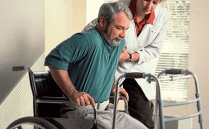
Signs of parietal lobes are often associated with somatic sensations, including:Sensory-tactile disturbances. Disturbances of body perception, postural sense, and passive motor sense. Sensory and visual distraction syndromes, loss of sensory and spatial attention. The patient may come to a state of denial of the existence of a limb. Loss of ability to read, write, and calculate. Inability to find a specific geographical location. Loss of ability to identify objects based on touch.
1.3 Signs of the temporal lobes
The temporal lobe signs are mainly related to memory and hearing, including:Deafness not due to damage to structures in the ear, also called cortical deafness. Tinnitus, auditory hallucinations. Loss of the ability to understand music and language is called sensory aphasia. Amnesia (affects near memory, distant memory, or both). Memory Disorders Hallucinations are complex and varied. Partial and complex seizures (temporal lobe epilepsy).

1.4 Signs of the occipital lobe
Signs of common occipital lobes related to vision include:Total loss of vision (cortical blindness). Loss of sight but the patient denies that he is lost (Anton syndrome). Unilateral visual acuity loss in both eyes (ipsilateral hemiplegia). Visual impairment, for example, loss of the ability to recognize familiar objects, colors or faces. Illusion as seeing things smaller or larger. Visual hallucinations, manifesting as basic shapes, such as zigzags or flashes of light, in one half of the visual field in each eye. In contrast, visual hallucinations of the temporal lobe take on complex forms and occupy the entire field of vision.
1.5 Signs of cerebellum
Cerebellar signs, often related to balance and coordination, may include:Clumsy and uncertain movements of the trunk and extremities (ataxia). Loss of coordination of subtle movements (intention tremor), eg finger-pointing in the nose-to-finger-pointing test. Fast-changing movements are not possible, for example, not being able to turn the hand over and over very quickly. Presence of involuntary nystagmus (Nystagmus).
1.6 Signs of the brain stem (Brain Stem)
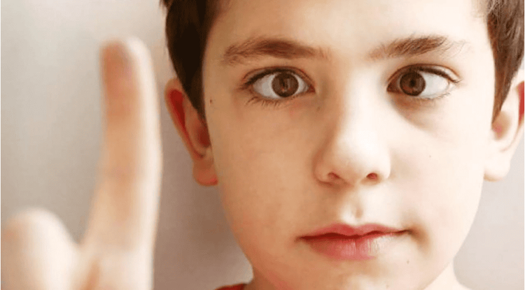
Brainstem signs may be due to specific sensory and motor disturbances, depending on the nerve and cranial nerve nuclei involved.Weakness, paralysis or sensory disturbance in extremities, crossed sign (with signs of injury in the face and trunk on the opposite side). Postural ataxia (loss of postural balance when the patient is upright, with head and body movements). In addition, the patient also has difficulty speaking, swallowing, eyeball twitching, and conjunctival vision.
1.7 Signs of the spinal cord
Spinal cord findings usually present with ipsilateral paralysis and loss of pain sensation on the contralateral side.2. Examine focal neurological signs
The examination of focal neurological signs is mainly based on signs to make a diagnosis. In addition, the doctor may examine:Physical examination
If the patient is awake: Do the Baré arm maneuver, Raimist maneuver and Mingazini maneuver. If the arm or leg is paralyzed, it can't be done or it's very weak and difficult. If the patient is comatose: Observe when the patient stretches out on the side of the body that is paralyzed or weak, the limbs on that side will move less or not at all. Meanwhile, the opposite half of the body, the non-paralyzed side, the limbs contract and struggle. Pain examination: Use a needle or pinch the patient's chest or inner arm to see which side responds to painful stimuli more clearly. Often relieves pain on the same side as the paralyzed side.
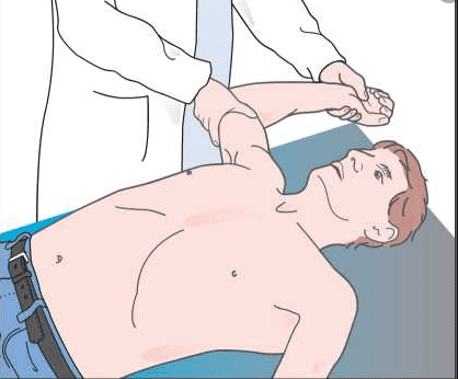
Examination of tendon reflexes Examination of the plantar reflexes (Babinski sign) Examination of the cranial nerves
Examination of pupillary reflexes to light
Focal neurological signs are classified into separate groups , showing cognitive and behavioral manifestations. Based on that, the doctor can accurately diagnose the focal neurological disease and give the most appropriate treatment.
Any questions that need to be answered by a specialist doctor as well as if you have a need for examination and treatment at Vinmec International General Hospital, please book an appointment on the website to be served.
Please dial HOTLINE for more information or register for an appointment HERE. Download MyVinmec app to make appointments faster and to manage your bookings easily.


