This is an automatically translated article.
The article was professionally consulted by Specialist Doctor I Le Hong Lien - Department of Obstetrics and Gynecology - Vinmec Central Park International General Hospital.1. Fetal ultrasound
One of the most widely used ultrasound applications is fetal ultrasound. Fetal ultrasound is a method that uses high-frequency sound waves to record images of the baby as well as the mother's genital organs such as uterus, ovaries, etc., in order to regularly monitor the development of the fetus. of the fetus as well as detecting abnormalities of the fetus or abnormalities of the mother so that early intervention can be given to minimize the impact.The average number of ultrasound scans can vary from pregnancy to period. Along with conventional ultrasound, some more advanced types of ultrasound are also gradually being applied such as color ultrasound, 3D ultrasound, 4D ultrasound and fetal echocardiography.
So what to do with fetal ultrasound? Fetal ultrasound may be used for several reasons. One of them is to monitor the development of the fetus and mother. In addition, the doctor may order a pregnancy ultrasound when detecting problems in a previous ultrasound or blood test. Ultrasound can also be performed for simpler purposes such as parents wanting to regularly see pictures of their baby before the baby is officially born or in some cases ultrasound to determine the sex of the fetus. . However, the above reasons are generally not recommended, especially for ultrasound to determine the sex of the fetus.

2. Some advanced fetal ultrasound techniques are often applied
Advanced fetal ultrasound techniques can make ultrasound images clearer and more detailed. They provide doctors with the information they need to make accurate diagnoses. Some commonly used ultrasound techniques:Transvaginal ultrasound : This technique helps to provide a clearer picture of the fetus, often used in the early stages of pregnancy when other types of ultrasound do not can provide clear images. This method uses an ultrasound probe inserted into the mother's vagina and the resulting images are displayed on a monitor. 3D ultrasound: Unlike traditional 2D ultrasound, 3D ultrasound provides a more general picture to assess both width, height and depth of the fetus. The procedures for performing 3D ultrasounds are similar to those for other types of ultrasound but using special transducers and software to create 3D images. This method also requires the technicians performing ultrasound to be thoroughly trained, so in practice it is rarely used. 4D ultrasound is a special form of 3D ultrasound, but the results of 4D ultrasound are created out a moving video of the fetus. This helps doctors as well as parents of the baby can better see the baby's face and movements.

3. Purpose of fetal ultrasound
Depending on the time of pregnancy, fetal ultrasound may be indicated with different purposes such as:In the first 3 months, fetal ultrasound is performed to:
Confirm the mother's pregnancy, determine the gestational age and estimated due date Check fetal heart rate, detect abnormalities if any Check possible abnormalities of placenta, uterus, ovaries... Diagnosis of ectopic pregnancy or risk miscarriage During the second and third trimesters of pregnancy :
Monitor the growth and position of the fetus Determine the sex of the baby Check the placenta for cervical occlusion or placental abruption immature or not. Check for features of Down syndrome (usually done between 13-14 weeks gestation). Check for birth defects or abnormalities in the fetus's structure or blood flow, to determine if the fetus is getting enough oxygen. Diagnosing problems with the mother's ovaries or uterus. Measure the length of the cervix, measure the hole in the cervix to screen for the risk of preterm birth, or guide other techniques such as amniocentesis. Confirm fetal death.

4. Pregnancy ultrasound procedure
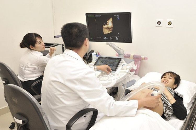
The non-invasive fetal ultrasound procedure is done in about 30 minutes to 1 hour with relatively simple steps:First, the technician asks the mother to take off her clothes and jewelry, put on the patient's shirt and lying on the hospital bed The mother will then be asked to lift her shirt to reveal her abdomen and pelvis. The technician will proceed to gently apply a layer of gel to the area to be ultrasounded to make sure there is no space between the transducer and the mother's skin. Next, using a transducer, it is gently moved over the mother's abdomen until an image of the fetus is captured on the monitor. Then check if the image quality is clear enough and if satisfactory, they will wipe off the gel to finish the ultrasound process. For transvaginal ultrasound, the only difference is that instead of moving the transducer over the mother's abdomen, the technician will use the transducer inserted into the vagina to take pictures of the fetus.
Fetal ultrasound is the leading popular application of the ultrasound method. Although not recommended for use to determine the sex of the fetus, fetal ultrasound can help parents get the first impression of the fetus in the womb, and can also monitor the development of the fetus. and promptly handle if abnormalities are detected. Today, with the development of science and technology, the quality of fetal ultrasound is also constantly improved and enhanced, from which parents have a clear image of the fetus.
Vinmec International General Hospital system uses the most modern generations of color ultrasound machines today in examining and treating patients. In addition, a team of experienced doctors and nurses will greatly assist in the diagnosis and early detection of abnormal signs of the body in order to provide timely treatment.
Specialist I Le Hong Lien has been an obstetrician-gynecologist at Vinmec Central Park International Hospital since November 2016. Doctor Lien has over 10 years of experience as a radiologist in the Department of Ultrasound at the leading hospital in the field of obstetrics and gynecology in the South - Tu Du Hospital.
Reference source: healthline.com; marchofdimes.org
Please dial HOTLINE for more information or register for an appointment HERE. Download MyVinmec app to make appointments faster and to manage your bookings easily.


