This is an automatically translated article.
Anterior chamber angle is a method often performed during routine eye exams, aiming to detect the risk of glaucoma. From there, the doctor can make a timely treatment plan for the patient, reducing the risk of complications of glaucoma.
1. What is the front corner?
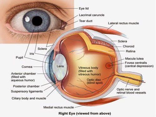
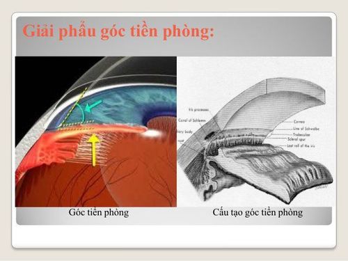
Anterior chamber is the space measured from the back of the cornea to the front of the iris. Anterior chamber angle is an acute angle formed by the cornea - iris. The apex of the anterior chamber angle corresponds to the outer corneal-sclera margin. In the corner of the front room, there are two important structures, namely the Schlemm tube located in the raft net area.
Front room angle is distinguished as follows:
Wide angle: ≥ 45°; Narrow angle: 20° - 45°; Closing angle: 0° - 20°.
2. What is the front corner of the room?
Anterior chamber angle examination is a method used to examine the eyes and assess the condition of the anterior chamber. During this procedure, your doctor will use glasses to look into the anterior angle of the room (the angle in the front part of the eye made up of the cornea and the iris).
Anterior chamber angle examination is a painless procedure, performed to check whether the anterior chamber angle is open or closed, and wide enough for fluid in the eye to circulate. This is usually done during a routine eye exam, depending on your age and risk of glaucoma.
3. The meaning of looking at the front corner of the room
Do it if your doctor suspects glaucoma. Glaucoma is a facial disease that can damage the optic nerve, causing blindness; Check the outlet angle of the closed or near-closed eye to determine the type of glaucoma you have; Detect scratches or some other damage related to the exit angle of the eye; Glaucoma treatment: When taking anterior chamber angle, the laser is directed to the anterior chamber angle through a special lens, which helps to reduce pressure in the eye and control glaucoma; Check for birth defects that cause glaucoma. Prophylaxis is contraindicated in cases of systemic disease that are not yet allowed to be performed.
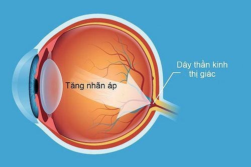
Soi góc tiền phòng hỗ trợ điều trị tăng nhãn áp
4. Perform a corner scan of the front room
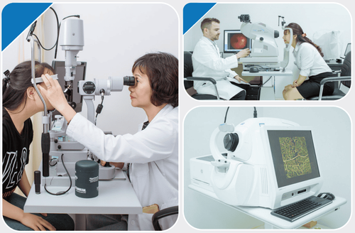
4.1 Preparation of Personnel: Ophthalmologist; Means: Pupil dilator, ophthalmic microscope and 3-mirror glasses; Patient: The purpose and procedure were explained, blood pressure history checked, contact lenses removed before the test (if any) and not put back on for 1 hour after the test until the anesthetic wears off; Medical records: In accordance with regulations of the Ministry of Health. 4.2 Procedures Checking records; Examination of the patient; Technical implementation: Using topical anesthetic Dicaine eye drops; The doctor sits facing the patient; The patient rests his chin on the microscope for eye examination; Place the 3-sided mirror glass in contact with the patient's cornea, observe at the smallest glass and move the microscope optical system forward until the anterior corner of the room is visible.
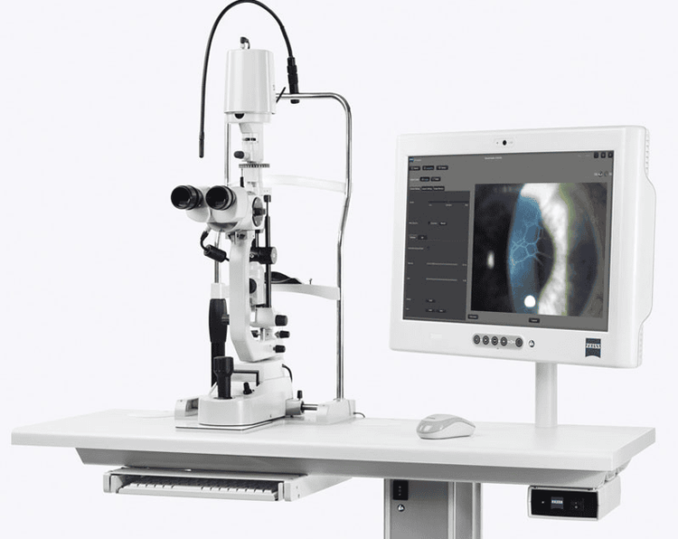
Máy sinh hiển vi khám mắt
The process of scanning the anterior chamber takes about 5 minutes.
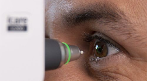
4.3 Post-procedural follow-up After instillation, patients may experience blurred vision for several hours after anterior angulation. Patients should not rub their eyes for 20 minutes after the test and it is best not to rub their eyes until the medication wears off. If suspecting that the patient has glaucoma, the doctor will order some more tests to measure the eye pressure, check the optic nerve,...
Evaluate the patient's condition, monitor the detection of these conditions. abnormal signs to report to the doctor for timely treatment. This test method has almost no side effects.
5. How to read the results of scanning the front corner of the room
Normal results: Anterior chamber angle in the eye is normal, enlarged, and unobstructed; Abnormal results: Anterior chamber angle is narrow, inserted or blocked. This means that the eye corner is partially or completely blocked and later the anterior chamber is at risk of being closed. Anterior chamber angle is a method to help diagnose glaucoma or other eye diseases. When asked to perform this technique, patients need to strictly follow the doctor's instructions to get quick and accurate results, avoiding the risk of complications.
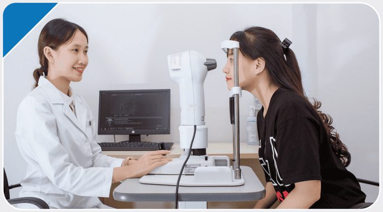
Người bệnh nên gặp bác sĩ khi gặp vấn đề về mắt
To meet the needs of patients for screening for eye diseases, Vinmec International Hospital currently provides Glaucoma Packages to help customers detect diseases early and have effective treatment, prevent dangerous complications.
Currently, Vinmec is implementing 2 packages of Glaucoma examination and treatment, including: Glaucoma early detection package and scleral trabeculectomy package to help:
Comprehensive eye examination and indication for surgery (if any) by experienced medical specialist. Advice on drugs, food, factors that may affect surgery.. Anesthesia examination and resuscitation assess the patient's general condition before surgery. Explain surgical prognosis.
To register for examination and treatment at Vinmec International General Hospital, you can contact Vinmec Health System nationwide, or register online HERE.
Recommended video:
Periodic health check at Vinmec: Protect yourself before it's too late!
LEARN MORE
Instructions on how to care for your eyes after Glaucoma Is the headache around the eyes dangerous? Glaucoma, glaucoma can be operated?


