This is an automatically translated article.
The article is written by Master, Doctor Nguyen Manh Ha - Radiotherapy Doctor - Radiation Therapy Center - Vinmec Times City International General Hospital.Chu Van Dung(1), Ha Ngoc Son)1), Nguyen Van Han(1), Nguyen Van Nam(1), Nguyen Trung Hieu(1), Pham Tuan Anh(1), Tran Ba Bach(2), Nguyen Dinh Long (2), Doan Trung Hiep (3), Nguyen Manh Ha (3).
(1) Technician; (2) Radiotherapy engineer, (3) Radiotherapy Doctor - Department of Radiation Therapy - Vinmec Times City General Hospital.
Email: v.dungcv@vinmec.com Mobile: (+84) 965 095 556
Summary
Objective: To compare dose levels for treatment volume and dose to critical organs between 3D techniques -CRT and VMAT in the NB phase of holding breath at the end of expiration in preoperative radiotherapy for esophageal cancer. Method of controlling beam dose to the treatment volume during irradiation by relying on daily setup errors to study the PTV margin opening for 4D technique.
Subjects and research methods: A total of 10 esophageal cancer patients were prescribed preoperative radiotherapy from January 2019 to April 2020 at Vinmec Times City International General Hospital. All 10 patients were scheduled to dose according to 2 techniques 3D-CRT and VMAT in the NB phase of end-expiratory apnea. All dose calculation parameters on DVH and 2D-KV setup errors on OBI software were analyzed.
Result: VMAT's CI is better than 3D-CRT (p=0.009), VMAT's HI index is lower than 3D-CRT but not statistically significant (p=0.205). Statistics of dose calculation on adjacent organs between 2 techniques: for lung V5Gy, V10Gy of 3D-CRT is lower than VMAT (P-value 0.01 and 0.02), respectively, while important indexes V20Gy, V30Gy of VMAT are better (p-values are 0.03 and 0.00, respectively). For heart V5Gy, V10Gy of 3D-CRT was lower than VMAT (P-value 0.014 and 0.03) but for V20Gy, V30Gy, V40Gy of VMAT lower than 3D-CRT (p 0.03, respectively, 0.01 and 0.00). The dose to the spinal cord of VMAT is also lower than 3D-CRT with statistical significance p = 0.00. Based on 230 pairs of 2D-KV images, the average setting error in 3 directions AP, SI, LR is (1.41 1.78mm) , (0.88 ± 2.69mm), 1.06± respectively. 1.36mm.
Conclusion: VMAT gives better results for calculating dose to PTV as well as reducing dose to critical organs such as heart, lungs, spinal cord better than 3D-CRT. The setting error according to Van-Herk in 3 directions AP, SI, LR is 4.8mm, 4.1mm, 3.6mm respectively.
Keywords: Esophageal cancer, breathing control system, dose comparison, setting error.
I. ASK THE PROBLEM
Esophageal cancer ranks 6th in incidence and 8th in mortality globally. In Vietnam, esophageal cancer is also a common cancer, ranking 12th in incidence and 9th in mortality for both sexes; but ranked 6th in incidence and 5th in male mortality [2]. Surgery is the most radical treatment for patients with esophageal cancer, but with unclear symptoms or signs of the disease, it is difficult to detect in the early stages, often when the tumor has progressed to a large size. Newly diagnosed and treated patients. Therefore, patients often undergo preoperative radiation therapy to reduce the size of the tumor and then start surgery. The combination of radiation therapy and surgery is very effective in the treatment of patients with esophageal cancer.In 2015, Stefan Münch et al. [6] published a study on preoperative radiotherapy for patients with esophageal cancer performed by 2 techniques 3D-CRT and VMAT with the results of patients being operated after radiation therapy. With treatment, averaging about 33 days for VMAT and 34 days for 3D-CRT, complete resection of the tumor was achieved 100% (VMAT) and 89.5% (3D-CRT) with a 3-year survival rate of 65% for VMAT and 45% for 3D.
The esophagus is a mobile organ by breathing, delivering sufficient dose to the tumor but reducing the radiation dose to neighboring organs is a big challenge in the treatment of esophageal cancer. Currently, with improvements in radiotherapy techniques: the combination of RPM (Real-Time position management) breathing control system with 3D-CRT and VMAT techniques to form 4D (four dimension) techniques. Current best technique in the treatment of UTI. From 2006, Lorchel F. JL Dumas et al. [4] to 2013 Guanzhong Gong et al. [1] both published studies showing that dose delivery to the tumor and reduced dose to the heart and lungs from Inspiratory end-radiation technique is better than free breathing in the treatment of esophageal cancer. However, we found that it is very difficult to keep the breath steady when the patient takes a deep breath, much worse than if the patient exhales and holds his breath. Holding the breath during deep inhalation is not guaranteed throughout the course of radiation therapy, which greatly affects the irradiation to the tumor.
Currently in Vietnam for radiation therapy for rectal cancer patients, the technique of end-expiratory radiotherapy is being applied to patients with colorectal cancer. Therefore, we carried out a study to evaluate the dose-level indices of the treatment volume and the radiation dose to the critical organs of patients with preoperative radiotherapy for esophageal cancer using 3D-CRT and VMAT radiotherapy techniques. control breathing at the end of expiration.
II. RESEARCH SUBJECTS AND METHODS
II.1 Subjects A total of 10 esophageal cancer patients were assigned preoperative radiation therapy through the Tumorboard from January 2019 to April 2020 at Vinmec Times City International General Hospital. . All 10 patients were scheduled for dosing according to 2 techniques 3D-CRT and VMAT at the end-expiratory cessation NB phase to compare and contrast dosing indices for treatment volume and organs. vicinity. Based on the 2D-KV images taken daily recorded above recorded on the OBI (on-board imaging) system compared with DRR (Digital Reconstruction Radiography) images to determine the setting error in all 3 direction.II.2 Study procedure II.2.1 Simulated CT scan with controlled breathing (BH: Breath Hold) The patient was placed in a supine position using a vaclock or a SBRT (Stereotactic body radiotin therapy) table. two hands raised above the head grasp the two columns of the wingboard (CIVCO, Orange City, IA). An RPM (Real-Time position management) breathing synchronization system including camera and marker block (Varian Medical systems, Palo Alto, CA) was used for the BH technique. Before the simulation, all patients were trained in breathing management procedures, with a minimum breathing time of 20 seconds. Simulated CT scan on Optima 580 (GE Medical System, Milwaukee, Wisconsin USA), 2.5mm slice thickness, cut from the base of the skull to the end of the liver. Two sets of CT images, one without contrast, are used for treatment planning, and the other with contrast injection is used for the physician to draw organs during planning. Both sequences were taken with the patient holding the breath at the end of expiration.
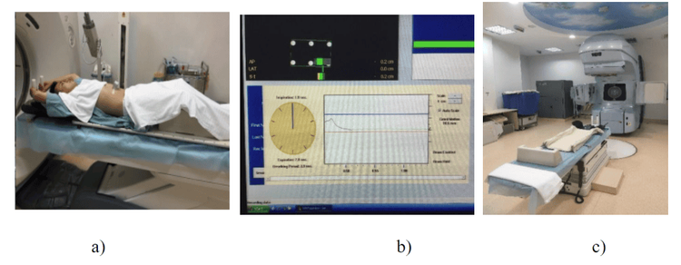
Hình 1: Đặt tư thế cố định bệnh nhân trong chụp CT (a), Tín hiệu nhịn thở của hệ thống RPM (b) và Bộ dụng cụ cố định của bệnh nhân (c).
II.2.3 Prescribing All 10 patients received radiation therapy with a total PTV dose of 41.4 Gy, divided dose 1.8Gy/1 day. The course of radiation therapy lasted 23 sessions, with 5 sessions per week.
II.2.4 Planning and approval of plans All 10 patients were planned on Eclips 13.0 software (Varian Medical System, Palo Alto, CA, USA) 2 3D-CRT plans for each patient. and VMAT, MLC 120 leaves.
II.2.4.1 Projection field design The 3D-CR,T plan is made with 4 projection fields with 15X photon energy levels, 2 AP-PA fields (0 degrees-180 degrees) designed with field engineering. In the field (FiF: Field-in-Field) to increase the uniformity of dose delivery in the treatment volume, reduce the hot dose (hotpost), 2 LR-RL fields are designed in an oblique direction (330 degrees-150 degrees) to avoid Maximum spinal cord ensures that the tolerated dose is strictly adhered to.
The VMAT plan is designed with 6 partial Arcs with angles of 181-231 degrees; 23-181 degrees; 39-309 degrees; 309-39 degrees; 179-103 degrees; 103-179 degrees, has the same isocenter with the 6X photon energy level. Because each time the patient holds his breath for 15-20 seconds, we have to subdivide the Arc into partial Arcs
II.2.4.2 Plan evaluation parameters Based on the dose-volume chart (DVH: Dose Volume Histogram), analyzed and calculated indexes: Dmax, Dmin, Dmean, CI (Comformity Index: homogeneity index), HI (Homogeneity Index: homogeneity index) were evaluated for the treatment volume (PTV). A high CI means that PTV is given a high dose of homogeneity and vice versa. A low HI indicates better dosimetry for PTV and vice versa. CI= V95%V x V95%TV95% (TV95% is the 95% isotopic volume, V: volume of PTV, V95%: volume of PTV receiving 95% dose). HI= D2%-D98%D50% (D2%, D50%, D98% are the doses that 2%, 50%, 98% of PTV received). Dmax, V5Gy, V10Gy, V20Gy, V30Gy, V40Gy for critical organs (OAR): Heart (heart), Lungs (lungs) and SpinalCord (spinal cord). In addition, the MU value comparison of 2 3D-CRT and VMAT plans is also recorded and compared.

Bảng 1: Tiêu chuẩn đánh giá kế hoạch xạ trị
II.3. Radiation dose control method In order to properly irradiate the tumor volume and reduce toxicity to neighboring organs, the border opening from CTV to PTV needs high accuracy. According to ICRU, the margin opening from CTV to PTV needs to take into account the accuracy of CTV position (IM: internal margin) as well as the accuracy of patient setting (SM: setup margin): CTV-PTVmargin=IM+SM. According to the van Herrk formula: SMvanHerk = 2.5 Σsetup +0.7 σsetup , in which, the setting error (SMvanHerk: setup margin), the systematic error (Σ: systematic error) is determined by the standard deviation of The mean setting errors for each patient, the random error (σ: random error) is determined by the mean of the standard deviations for each patient. Based on the comparison between 2D-kV images with DRR images according to bone landmarks. Total 230 sets of error settings in all 3 directions.

Hình 2: So sánh hình ảnh 2D-kV với hình ảnh DRR trước kho chiếu xạ.
III. RESULTS AND DISCUSSION
III.1. 3D-CRT plan and VMAT . plan
Hình 3: Thiết kế trường chiếu kế hoạch VMAT: 3 ARC (trái) và kế hoạch 3D-CRT: ha 1 (AP-PA), Pha 2 (RL-LR) tránh tủy hòan toàn (Field in Field)

Hình 4: Đường đồng liều 95% bao khít PTV trên kế hoạch VMAT (trái) và bao chùm ra ngoài PTV khá nhiều trên kế hoạch 3D-CRT (phải).

Bảng 2: Các thông số tính liều đối với PTV (TB: Giá trị trung bình, ĐLC: Độ lệch chuẩn).
III.3 Critical organ dose (OAR) parameters

Bảng 3: Thống kê liều vào các cơ quan nguy cấp

Hình 4: So sánh biểu đồ liều DVH của 2 kỹ thuật 3D-CRT và VMAT với PTV (màu xanh), Tim (a), Phổi (b) và Tủy sống (c).

Bảng 4: Sai số hệ thống (Σ), sai số ngẫu nhiên (σ) và sai số cài đặt (SM).

Biểu đồ 1: Biểu đồ phân bố sai số cài đặt trung bình theo từng bệnh nhân UTTQ.
IV. CONCLUSION
The parameters of dose-to-treatment volume using VMAT technique give better results than 3D-CRT technique, and VMAT also allows to reduce the dose of radiation to critical organs such as spinal cord, heart, and lungs.Preoperative rectal cancer radiotherapy using 3D-CRT technique with controlled breathing at the end of expiration phase at Vinmec Times City General Hospital, currently shows the setting error in 3 directions AP, SI, RL times turns are: 4.8mm, 4.1mm, 3.6mm. Thereby providing a suggestion for radiation therapists in opening the CTV-PTV border.
Please dial HOTLINE for more information or register for an appointment HERE. Download MyVinmec app to make appointments faster and to manage your bookings easily.
REFERENCESGuanzhong Gong (2013), Reduced lung dose during radiotherapy for thoracic esophageal carcinoma: VMAT combined with active breathing control for moderate DIBH, gong et al. Radiation oncology 2013,8:291. IARC: http://globocan.iarc.fr/default.aspx. Kaleigh Doke (2017) , Dosimetric effects of deep inspiration breath hold for radiation treatment of esophageal cancer based on tumor location, journal of clinical oncology.35(4_suppl):173-173. Lorchel F. JL Dumas (2006), Dosimetric consequences of breath-hold respiration in conformal radiotherapy of esophageal cancer, physica medical Vol XXII, N. 4 (October 2006) M.Dieters, Significant heart dose reduction by deep inspiration breath hold for RT of esophageal cancer , ESTRO36, P0-0070, s366 Stefan Münch (2015), Comparison of Dosimetric Parameters and Toxicity in Esophageal Cancer Patients Undergoing 3D Conformal Radiotherapy or VMAT, Springger-Verlag Berlin Heidelberg (2016)


