This is an automatically translated article.
The article is professionally consulted by Master, Doctor Nguyen Ngoc Thang - Gastroenterologist - General Surgery Department - Vinmec Danang International General Hospital.The esophagus is a muscular tube that connects the throat (pharynx) to the stomach. The esophagus is about 8 inches long, and is lined with moist pink tissue called the mucosa. The esophagus moves behind the windpipe (windpipe), the heart, and in front of the spine. Just before entering the stomach, the esophagus passes through the diaphragm.
1. What is the esophagus? Structure of the esophagus
The esophagus is a muscular tube that connects the throat (pharynx) to the stomach. The esophagus is about 8 inches long, and is lined with moist pink tissue called the mucosa, which is flattened because the walls press against each other. The esophagus is shaped like a tube when food is swallowed. The esophagus moves behind the windpipe (windpipe), the heart, and in front of the spine. Just before entering the stomach, the esophagus passes through the diaphragm. The esophagus is relatively mobile and adheres to the viscera by a loose structure.
In terms of surgery, the esophagus is divided into 3 segments:
Upper 1/3 of the esophagus: This segment is only about 10cm long, starting from the mouth of the esophagus to the upper border of the aortic arch. The middle third of the esophagus: This segment is about 8cm long, starting from the superior border of the aortic arch to the lower border of the inferior pulmonary vein. Lower 1/3 of esophagus: This segment is about 7cm long, starting from the lower border of the pulmonary veins to the cardia. Passes through the oesophagus with many blood vessels with blood supply varying with the course and length of the esophagus as follows:
The upper esophageal sphincter and the upper part of the esophagus receive blood from the glandular artery True thoracic segment The bronchial artery, which receives blood directly from the thoracic aorta, is the esophageal branch. Receives blood from the left gastric artery and the left lower pulmonary artery is the thoracic esophagus and the true sphincter. The upper esophagus (UES) is a bundle of muscles at the top of the esophagus. The muscles of the UES are consciously controlled, acting on breathing, eating, burping, and vomiting. UES keeps food and secretions out of the trachea.
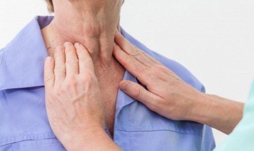
Thực quản là một ống cơ nối cổ họng (hầu họng) với dạ dày
The lower esophageal sphincter (LES) is a muscle bundle at the lower end of the esophagus, where the stomach meets. When the LES is closed, it prevents acid and stomach contents from traveling backwards from the stomach. The LES muscles are not controlled voluntarily.
In terms of structure: The layer of connective tissue that surrounds the esophagus is the outermost layer. Consists of 3 layers below the connective tissue layer of the esophagus. Specifically:
The muscle layer includes smooth muscle and striated muscle: In which, up to 2⁄3 of the lower esophagus is smooth muscle and 1⁄3 of the upper esophagus is skeletal muscle. The outer longitudinal bands and the inner circular muscle fibers are called smooth muscles. The bundles of striated muscle fibers, which surround the pharynx, become thinner downwards, and then reappear in the cardia to form the cardiac sphincter (lower esophageal sphincter) which is the striated muscle. The submucosa is composed of blood vessels and nerves. The mucosal layer is composed of the epithelial layer, lining the lumen of the esophagus, and is composed of the epithelial layer, the lining layer, the muscular layer, and the glands.
2. Operation of the esophagus
The function of the esophagus is to carry food from the throat to the stomach. When eating, along with the lifting of the larynx, the muscles in the throat contract, pushing food from the mouth down the esophagus. To receive a moderate amount of food, the muscles in the mouth of the esophagus relax. Food that is pasty or liquid will not fall into the stomach. Thanks to the slow peristaltic waves of the esophagus, combined with the weight of the food, denser foods move through the esophagus. Here, the main task of the esophagus is to push food to the heart and stomach:
To prevent air from entering the esophagus when it is open, the mouth of the esophagus is usually closed. When you do the swallowing action, it opens the Heart which is also normally closed. To prevent reflux of fluid, it acts as a one-way valve Mucositis will cause the upper sphincter to not close as often and lead to disturbances in cases of myasthenia gravis.
Thực quản là một ống cơ nối cổ họng (hầu họng) với dạ dày
3. Diseases related to the esophagus
The upper esophageal sphincter (UES) is a bundle of muscles at the top of the esophagus. The muscles of the UES are consciously controlled, acting on breathing, eating, burping, and vomiting. UES keeps food and secretions out of the trachea.
The lower esophageal sphincter (LES) is a bundle of muscles at the lower end of the esophagus, where it meets the stomach. When the LES is closed, it prevents acid and stomach contents from traveling backwards from the stomach. The LES muscles are not voluntary.
Heartburn: The LES does not close completely allowing acidic stomach contents to back up (reflux) into the esophagus. Reflux can cause heartburn, cough or hoarseness, or no symptoms at all. Gastroesophageal reflux disease (GERD): When reflux occurs frequently or causes discomfort, it is called gastroesophageal reflux disease (GERD). Esophagitis: Esophagitis can be caused by irritation (such as from reflux or radiation therapy) or infection. Barrett's esophagus: Frequent reflux of stomach acid irritates the esophagus, which can cause the lower part to change its structure. In some cases, Barrett's esophagus progresses to esophageal cancer. Esophageal ulcer: an ulcer that occurs in an area of the lining of the esophagus. This is usually caused by chronic reflux. Esophageal spasm: Narrowing of the esophagus. Chronic irritation from reflux is a common cause of esophageal stricture. Achalasia: A rare disease in which the lower esophageal sphincter does not work properly. Difficulty swallowing and vomiting food are symptoms of this condition. Esophageal cancer: Although very serious, esophageal cancer is not common. Risk factors for esophageal cancer include smoking, heavy drinking, and chronic reflux. Mallory-Weiss tear: Re-vomiting creates a tear in the lining of the esophagus. Esophageal bleeding into the stomach, often accompanied by hematemesis. Esophageal varices: In people with cirrhosis, veins in the esophagus can become stretched and bulge called varicose veins, these veins are prone to life-threatening bleeding. Esophageal ring (Schatzki ring): A benign tissue accumulation that commonly occurs in a ring around the lower end of the esophagus. Schatzki's rings usually cause no symptoms, but can make swallowing difficult. Esophageal network: A tissue buildup (similar to an esophageal ring) commonly occurs in the upper esophagus. Like rings, esophageal webs usually cause no symptoms. Plummer-Vinson syndrome: A condition that includes chronic iron-deficiency anemia, esophageal webs, and difficulty swallowing. Iron supplementation and dilation of the esophagus are treatments.
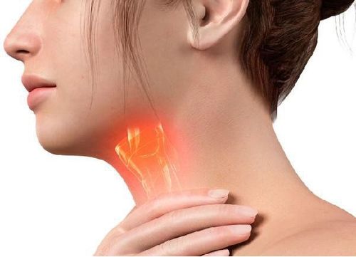
Tĩnh mạch trong thực quản có thể bị căng và phình ra
4. Diagnosis of diseases related to the esophagus
Here are some methods of diagnosing diseases related to the esophagus:
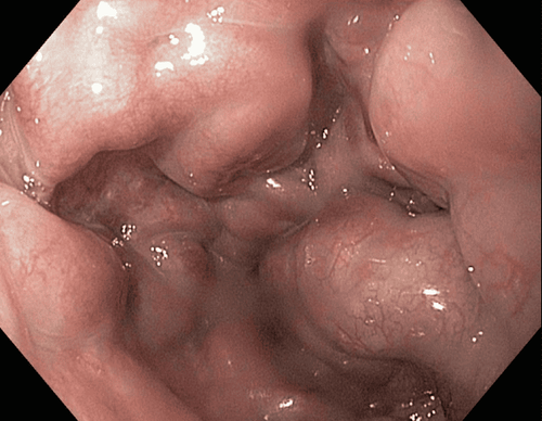
Upper endoscopy, EGD (esophagogastroduodenoscopy): A flexible catheter with a camera on its end (endoscope) is inserted through mouth. Endoscopy allows examination of the esophagus, stomach, and duodenum (small intestine). Esophageal pH monitoring: A probe that monitors acidity (pH) is inserted into the esophagus. pH monitoring can help identify GERD and monitor response to treatment. Scan with Barium: the patient will be given a solution called Barium or a coating containing Barium, then X-rays are used to take pictures of the esophagus and stomach. Usually, oral administration of a barium solution is used to look for the cause of dysphagia.
5. Treatment of diseases related to the esophagus
Several methods are applied to treat diseases related to the esophagus, including:
H2 blockers: Histamine stimulates the release of acid in the stomach. Certain antihistamines called H2 blockers can reduce acid and improve GERD and esophagitis. Proton pump inhibitors: These drugs block acid production in the stomach wall. Reducing stomach acid can relieve GERD symptoms, and help an ulcer or esophagitis heal. Esophageal resection: Surgery to remove the esophagus, usually applied to patients with esophageal cancer. Esophageal dilation: A balloon is passed down the esophagus and inflated to dilate a limit, web, or ring that interferes with swallowing. Esophageal vein ligation: During an endoscopy, rubber band-like devices may be wrapped around esophageal varices. Esophageal vascular ligation helps to reduce the possibility of bleeding. Biopsy: Usually done through an endoscope, a small piece of the esophagus is taken as a sample for evaluation under a microscope. Laser endoscopy: A new method of using a microscope to look inside a patient, which could replace the need for multiple biopsies.
Please dial HOTLINE for more information or register for an appointment HERE. Download MyVinmec app to make appointments faster and to manage your bookings easily.
Reference source: webmd.com