This is an automatically translated article.
Cranial basal tumor is a very difficult lesion to operate. Choosing the right surgical method can bring good results for the patient. Therefore, the method of endoscopic surgery to remove skull base tumors can be applied to many lesions.1. Purpose of skull base tumor surgery
The purpose of cranial basal tumor surgery is to remove the tumor as much as possible, while preserving important functions and reducing the rate of complications as low as possible.
Cranial basal tumor is a difficult lesion to operate. Choosing the right surgical method can bring good results for the patient. Therefore, the method of endoscopic surgery to remove skull base tumors can be applied to many lesions.
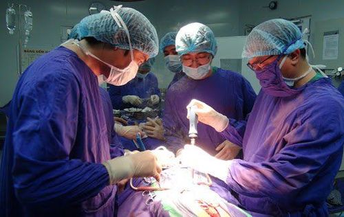
Phẫu thuật u nền sọ giúp loại bỏ khối u
2. Indications and contraindications for endoscopic skull base tumor surgery
Indications for endoscopic surgery to remove skull base tumor when: Craniopharyngeal tumor; anterior craniocervical meningioma; raw tumor; mucinous tumor; metastatic tumor; Cranial basilar neuroma is contraindicated when: This surgical method has no absolute contraindications, only relative contraindications for craniopharyngeal tumors located outside the carotid artery (approximately more than 10mm). ), or at locations far from the nasopharyngeal region; Vascular tumors are at risk of excessive bleeding.
3. Steps to perform skull base tumor surgery
Step 1: Prepare equipment for endoscopic brain tumor surgery including: Transnasal sinus surgery kit, pituitary surgical instruments, high-speed diamond drilling system, Navigation system, hemostasis instruments, skull base closure supplies, endoscopy system with camera and monitor Step 2: Clinical examination, specialized examination of ophthalmology, endocrinology, otorhinolaryngology; MRI scan of the brain, CT film evaluates the structure of the base of the skull. Step 3: Ask the patient to lie in supine position, head tilted to the surgeon 200, put gauze impregnated with naphazolin 2% 10 minutes before surgery. If the skull base must be enlarged, use the technique of closing the base of the skull with fascia, femoral fat, and nasal septal flap with palatal sphenoid vein. Step 4: Push the middle roll to the side to find the sphenoid sinus opening to open the sphenoid sinus, create a flap of the nasal septum with a feeding vessel, take a part of the nasal septum, open the anterior wall of the sphenoid sinus with Kerrison and drill. Step 5: Burn the mucosa at the opening of the bone, use a diamond drill to gradually grind the bone. Open the sclera slowly. Use diamond drill to stop bone bleeding. Step 6: Use a curette and a suction tube to remove the base of the skull and use 300, 700 optic to see the angles. Step 7: Use scale, thigh fat, septal bone fragment or septal flap with vascular pedicle; use bio-glue Bio Glue, Tisseel to create adhesive; put a Foley sonde to keep the graft for 34 days.

Phẫu thuật nội soi lấy u nền cần thực hiện ở bệnh viện lớn, có uy tín
Vinmec International General Hospital with a system of modern facilities, medical equipment and a team of experts and doctors with many years of experience in neurological examination and treatment, patients can completely peace of mind for examination and treatment at the Hospital.
To register for examination and treatment at Vinmec International General Hospital, you can contact Vinmec Health System nationwide, or register online HERE.
MORE
Is mediastinal tumor dangerous? Mediastinal tumor removal by open surgery How to treat mediastinal tumor?


