This is an automatically translated article.
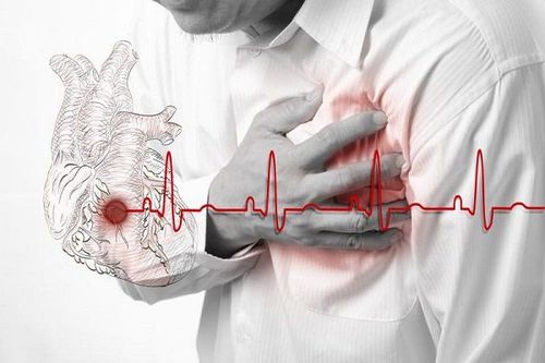
The article was professionally consulted by Specialist II, Senior Doctor Doan Du Dat - General Internal Medicine Doctor - Department of Medical Examination and Internal Medicine - Vinmec Ha Long International Hospital.Acute pericardial effusion is a rare disease, the frequency of which is only about 0.1% of the total number of hospitalized patients and about 5% of the total number of patients admitted to the emergency room because of chest pain. However, this is a very dangerous disease, so timely emergency pericardial effusion will help patients have a better prognosis.
1. Structure of the pericardium
The pericardium (pericardium) consists of the pericardium and the pericardium, which normally contains 10-50ml of fluid between these two layers. Pericardial fluid is absorbed by the thoracic lymphatic system.The pericardium helps to limit the excessive dilation of the heart chambers. Besides, the pericardium also helps keep the heart in a stable position in the ribcage, making the heart work smoothly, limiting friction and being a barrier to bacteria.
Màng ngoài tim có vai trò quan trọng với quả tim
2. What is pericardial effusion?
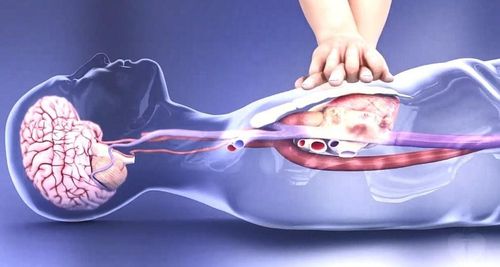
Normally, our heart is surrounded by a sheath and creates a pericardial cavity that contains fluid. When the condition of fluid in the pericardium increases, it will cause acute cardiac tamponade (the heart is like swimming in a pool of water), the chambers of the heart cannot expand, the blood cannot return to the heart, and the heart does not contract effectively. fruit. This leads to a drop in blood pressure and can cause death if the patient is not treated promptly.3. Causes of pericardial effusion
The causes of pericardial effusion are as follows:Pericarditis reaction: bacterial and viral infections; Pericardial effusion occurs when the outflow of pericardial fluid is blocked, and blood accumulates in the pericardium; Heart failure ; Coronary artery disease causing myocardial infarction with complications; Hypothyroidism ; Lupus erythematosus ; Rheumatoid arthritis ;

Suy giáp là một trong những nguyên nhân gây tràn dịch màng tim
4. Manifestations of pericardial effusion
4.1. Pericardial effusion without cardiac compression
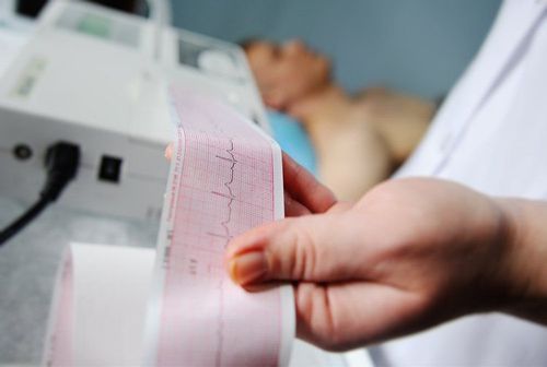
Pericardial effusion is usually asymptomatic or sometimes patients only have a dull ache, a feeling of heaviness in the chest, chest pain behind the sternum, pain that increases with deep breathing, and eases when the patient lies or sits down. person forward. However, if the pericardial fluid is small, the symptoms are often difficult to see, if the fluid is large, you will see signs such as hearing faint heart sounds, Edward's sign (cloudy percussion, bronchial murmur) and rales. due to secondary compression. Laboratory examination should be performed to diagnose pericardial effusion without cardiac compression:Electrocardiogram (ECG): diffuse low voltage, alternating electrocardiogram in case of large pericardial effusion. Cardiopulmonary imaging: The heart shadow is unchanged when the fluid is 1 to 2mm thick, the heart is enlarged when the pericardial fluid is more than 250ml. Echocardiography: this is the most effective method to diagnose pericardial effusion Other tests: transesophageal ultrasound, computed tomography, magnetic resonance imaging. Test of pericardial fluid: in case of a lot of fluid with aspiration, it is necessary to look for tubercle bacilli, biochemical, bacteriological and cytological tests.

Siêu âm tim giúp chẩn đoán tình trạng tràn dịch màng ngoài tim
4.2. Pericardial effusion with signs of cardiac tamponade
Low cardiac output: manifests as restlessness, anxiety or irritability, lethargy or possibly fainting, decreased urine volume, dyspnea, chest compression, loss of appetite and weight loss with chronic pericardial effusion. Increased central venous pressure, tachycardia, tachypnea, usually if the amount of fluid in the pericardial cavity is high, causing cardiac compression, often no pericardial rubbing is heard, mainly faint heart sounds, even no heart sound is heard. Symptoms similar to right heart failure: hepatomegaly, distended neck veins, pleural effusion. Hypotension, inversion (blood pressure lower than 10 mmHg on deep inspiration). Signs of tamponade include:There is fluid in the pericardial cavity (space on echocardiography). Inferior vena cava dilatation is more than 50% during deep inspiration, which has a sensitivity of 97% but a specificity of only 40% for the diagnosis of chest compressions. False left ventricle hypertrophy. In summary, the typical clinical symptom is the BECK triad: distended neck veins, low blood pressure and faint heart sounds. Laboratory examination to diagnose pericardial effusion with signs of cardiac compression:
Transthoracic echocardiography: mandatory when cardiac tamponade is suspected Right heart catheterization: applied in places where cardiac catheterization is performed card.
5. How to treat pericardial effusion?
5.1. Pericardial effusion without cardiac compression
When pericardial effusion is confirmed without signs of compression, it is necessary to treat the etiology and control the hemodynamic fluctuations caused by the pericardial fluid. Indications for percutaneous pericardial drainage in cancer, bacterial and fungal infections. Note, pericardial puncture should not be performed in cases of small pericardial fluid.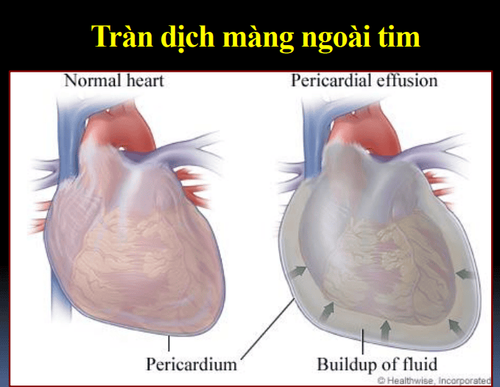
Bệnh nhân tràn dịch màng ngoài tim không có dấu ép tim được chỉ định chọc dẫn lưu dịch màng ngoài tim qua da
5.2. Pericardial effusion with signs of cardiac tamponade
Patients need to be hospitalized and receive rapid percutaneous pericardial drainage for decompression in emergency conditions, which is less invasive than other methods and requires minimal preparation. minimal. Aspiration drainage is the only treatment that needs to be done quickly. Conduct medical treatment by rehydration of electrolytes, use of drugs to raise blood pressure if the patient has hypotension such as: Norepinephrine, Dobutamine... should avoid vasodilators such as Nitroglycerine, Nitroprusside... Nong Percutaneous balloon pericardium (interventional cardiology): used when the emergency physician has extensive experience and in cancer patients causing pericardial effusion. Surgical drainage of pericardial fluid (if the medical facility has a heart surgery center): applied in cases of complicated pericardial effusion, postoperative or recurrent effusion...6. Pericardial drainage aspiration
Pericardial drainage aspiration is an active treatment method, which needs to be done quickly and with the right technique, ensuring patient life and minimizing complications.6.1. Prepare to suck
Fully prepare emergency equipment Place an intravenous line for the patient Monitor: electrocardiogram, SpO2 ... Anesthesia (in excited patients...) Atropin is injected to the patient Ultrasound control immediately before pericardiocentesis. Insert a nasogastric tube if the stomach is distended. Disinfect the puncture site with Povidine 10% Anesthetize the puncture site with Lidocaine 1%.
Trong trường hợp người bệnh bị kích động cần được gây mê
6.2. Posing pose
The patient lies in the Fowler position (45 degrees from the horizontal).6.3. Complications of pericardiocentesis
The incidence is about 4-40%, including:Arrhythmia Coronary artery puncture Left internal mammary artery puncture Hemothorax Pneumothorax Pneumopericardium Liver injury Thrombotic needle occlusion No suction fluid is obtained through the needle tip in the pericardial cavity. Heart chamber puncture: blood clots. Pericardial effusion is rare, but if not examined and treated promptly can leave dangerous complications, even death. Therefore, when there are signs of pericardial effusion, patients need to go to medical centers for examination and treatment.
If you have pericardial effusion. You can go to Vinmec International General Hospital for examination and treatment. Here, a team of qualified, experienced and well-trained doctors and nurses will examine, diagnose and advise patients with advanced and modern methods; Modern, advanced technology equipment ensures the most accurate results. Not only solve the cardiac tamponade due to effusion, but also diagnose the cause of pericardial effusion with the most accurate results. You will be satisfied with the results of our treatment.
Please dial HOTLINE for more information or register for an appointment HERE. Download MyVinmec app to make appointments faster and to manage your bookings easily.


