This is an automatically translated article.
The article is written by MSc.BS Mai Vien Phuong - Gastroenterologist, Department of Medical Examination & Internal Medicine - Vinmec Central Park International General Hospital.
Stomach cancer can be completely cured if detected and treated at an early stage. Therefore, early detection of the disease is very important. In Japan, Korea and some Western countries, the application of modern endoscopes such as narrow-band magnifying endoscopy (NBI) for early gastric cancer diagnosis is widely deployed, thus contributing to the development of gastric cancer. improve early gastric cancer detection rates.
In many countries around the world, stomach cancer is mostly diagnosed at a late stage, the main means of diagnosis in these places is a conventional endoscope under white light (i.e. endoscopy with mode). normal white light machine, do not use advanced modes such as staining endoscope, magnifying endoscope).
Widespread adoption of modern means for early cancer diagnosis in developing countries presents a major technical and economic challenge. Fortunately, conventional endoscopy can still detect early gastric cancer lesions if we proceed according to a standard.
Therefore, Japanese experts have issued Guidelines on the procedure of early gastric cancer diagnosis by conventional endoscopy. To do this, there are a few things to keep in mind.
1. Prepare your stomach well
Standard stomach preparation is a must, which helps to reduce the time and effort of doctors and technicians during the procedure, for example, to clean mucus and foam on the mucosal surface. gastric mucosa.
Patients need to fast for at least 8 hours before the procedure, foaming and mucolytic solutions are also used orally 30 minutes before the procedure. Indeed, small, flat lesions will sometimes be covered in leftover food or excessive foam in the stomach.

Người bệnh cần nhịn ăn trước khi tiến hành nội soi dạ dày
2. Assess the risk of stomach cancer as soon as the endoscope enters the stomach
As soon as the endoscope is inserted into the stomach, on the endoscopic image, the doctor needs to determine the risk factors for stomach cancer on the mucosal background (such as gastritis caused by Helicobacter pylori (HP) infection, atrophic gastritis. or intestinal metaplasia). If the mucosal background is normal, there are few lesions that suggest gastric cancer.
Gastric mucosa at low risk of gastric cancer : A unique finding is referred to as RAC (regular arrangement of collecting venules) on endoscopy. By closely observing the endoscopic image of the normal gastric body, small spider-like blood vessels can be seen. If these converging capillaries are regularly arranged, the sensitivity and specificity for the diagnosis of normal gastric corpus callosum not infected with H. If the stomach is not infected with H.Pylori, it is less likely to get stomach cancer. Gastric mucosa at risk of gastric cancer: Vascular imaging is clearly observed and the loss of mucosal folds in the gastric body is characteristic of chronic atrophic gastritis, a risk factor. of gastric cancer.
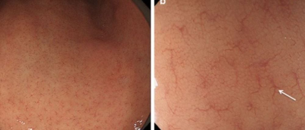
Hình 1: Hình ảnh niêm mạc dạ dày không nhiễm H.Pylori: Sự sắp xếp đều đặn của mao mạch hội tụ (RAC) trên nền niêm mạc dạ dày bình thường. Các mạch máu giống mạng nhện nhỏ tương ứng với sự sắp xếp các mao mạch đều đặn (mũi tên), ít có khả năng nhiễm H.Pylori ở niêm mạc dạ dày này.
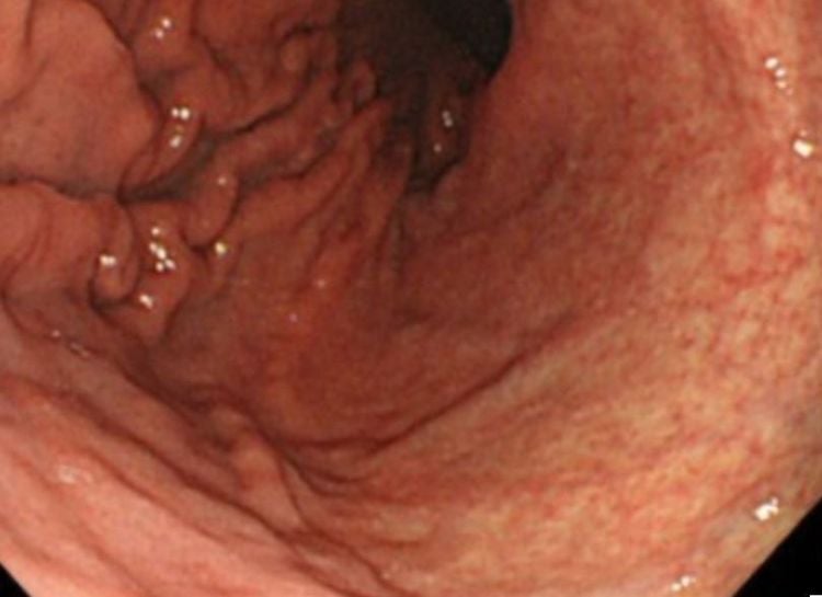
Hình 2: Vùng bên phải của hình ảnh: Niêm mạc dạ dày có viêm teo mạn tính do nhiễm H.Pylori, nhìn thấy được các mạch máu và mất nếp niêm mạc thân vị dạ dày trên nền viêm teo dạ dày mạn tính.
3. Find and detect early cancerous lesions
3.1 To detect lesions The first thing is that the doctor needs to be proficient in the sense of identifying suspected lesions. The most common signs: surface changes and color changes. Other findings, such as abrupt changes in the vascular/mucosa background, changes in light reflex, and spontaneous bleeding of the lesion.
Early gastric cancer with ulceration and polyps is easy to detect, just based on surface changes, provided that the stomach is fully visualized and the stomach is carefully prepared before surgery. tips. The endoscopist especially needs to watch out for these important signs in order to detect early gastric cancer that mimics gastritis.
The first important sign is surface change. Because this lesion is very small and has only a subtle change, it has an image similar to erosive gastritis, so it is difficult to detect, and it is easy to miss. However, it is important to be aware that such subtle changes can be an important sign of early stomach cancer. The second important sign is color change. A well-demarcated color change area is an important sign of gastric cancerous lesions resembling gastritis. Against the background of atrophic mucosa, the vascular background is clearly visible in the deep mucosal and submucosal parts. If the vascular background is traced, a well-demarcated area where the surrounding blood vessels are suddenly lost will be detected. This is a useful marker for detecting early cancerous lesions.
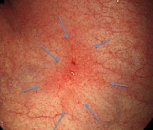
Hình 3: Một tổn thương ung thư dạ dày giai đoạn sớm, ở hang vị, với các dấu hiệu: tổn thương màu đỏ, lõm bề mặt, ranh giới rõ, có nền mạch máu niêm mạc xung quanh bị biến mất đột đột. Tổn thương này rất giống một tổn thương viêm dạ dày.
3.2 Lesions Identification To identify lesions, two main signs, color and surface morphology, are applied to the description on conventional endoscopy. The differential diagnosis is based on the following criteria:
Well-demarcated lesion Color or surface abnormalities If conventional white light endoscopy and/or staining endoscopy meet both of the above criteria , be careful, it is possible that this lesion is early stage stomach cancer.
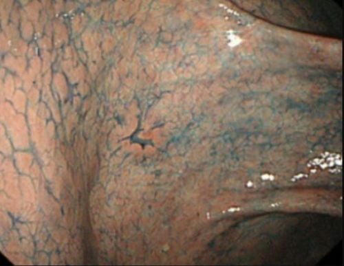
Hình 4: Tổn thương ung thư sớm rất nhỏ, có hình ảnh giống viêm dạ dày trợt, rất dễ bị bỏ qua.
Until now, the world is still increasing training to improve the ability to detect early gastric cancer on white light endoscopy alone. Early cancer detection skills, focusing on 3 basic things: technique, knowledge, and experience.
For these, the endoscopic surgeon can gain knowledge and techniques through attending regular lectures or seminars, training courses, and simple but repeated practice. Repetition will be helpful for maintaining ability.
“We only find what we know”... Good practice based on good technique, good knowledge and extensive experience will increase the doctor's ability to detect stomach cancer early. endoscopy as well as other early cancers.
Vinmec Central Park is equipped with the most modern digestive endoscope system of Olympus, one of the best brands today, including machines being treated like the Olympus CLV 190 endoscope. , with a team of doctors with extensive experience in early cancer diagnosis of the gastrointestinal tract, trained through many domestic and foreign training courses on early cancer diagnosis and treatment.
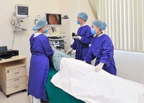
Khách hàng sử dụng dịch vụ nội soi dạ dày tại Vinmec
The advantage of these endoscopes is that in addition to the conventional white light endoscope, the system also has the function of narrow band of light (NBI), magnified endoscope, with high resolution NBI endoscopic images and High contrast makes it easier to detect and screen and diagnose gastrointestinal cancer at an early stage. Once the cancer is detected at an early stage, depending on the nature of the lesion, it will be cut by mucosal resection (EMR), or submucosal dissection (ESD) through flexible bronchoscopy, without having to undergo surgery.
Not only that, to ensure sterility of endoscopes, the hospital is also equipped with an automatic endoscope washing machine and RO water filtration system.
Customers can go directly to Vinmec Central Park to visit or contact hotline 0283 6221 166 for support.


