This is an automatically translated article.
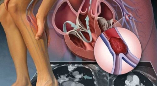
Deep vein ultrasound of the lower extremities is a non-invasive subclinical technique that helps diagnose a number of diseases related to the deep vein system of the lower extremities, the most important of which is venous thrombosis. It is known that this is a disease that brings many high-risk patients.1. Ultrasound of the deep veins of the lower extremities
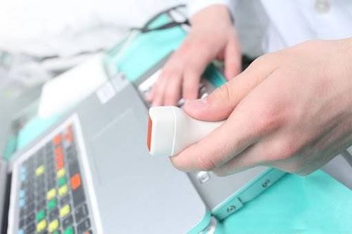
Vascular ultrasound of the lower extremities includes the arteries of the lower extremities and the veins of the lower extremities, including the superficial and deep veins. In particular, deep vein ultrasound of the lower extremities helps to diagnose and localize the damage to the deep vein system of the lower extremities, typically in cases of deep vein thrombosis of the lower extremities.To perform this technique, it is necessary to equip a Doppler ultrasound machine with a 7.5MHz or 5.0MHz type transducer capable of recording a low blood flow velocity of about 6cm/s, and at the same time, its high resolution should help people Performing ultrasound can survey and observe the veins located deep under the skin.
Regarding patient preparation, the patient will be asked to lie on his back, sit or stand depending on the goal of the ultrasound technique as well as the clinical presentation of the patient with symptoms of varicose veins, depigmentation, lower extremity edema or edema. In any position of skin ulcer, there will be suitable positions to perform ultrasound.

Siêu âm mạch máu chi dưới là một kỹ thuật cận lâm sàng
Bilateral ultrasound to compare and compare Comprehensive ultrasound from the inferior vena cava to other veins. When doing venous ultrasound, it also examines the subcutaneous tissues as well as the blood vessels in the vicinity. Ultrasonography in short axial view, cross-sectional anterior vasculature following the venous path to facilitate manoeuvrability. For lower extremity vein ultrasound, it includes the superficial and deep veins:
The superficial venous system includes the great saphenous veins and the small saphenous veins. The deep vein system of the lower extremities includes the common femoral vein, the deep femoral vein, the superficial femoral vein, the popliteal vein, the posterior tibial vein, the peroneal vein, the anterior tibial vein, the ventral leg vein, the sinus vein, and the vena cava. shoe muscle circuit. Normally, venous blood flow is so slow that ultrasound will not show Doppler signals in the peripheral veins, so performing hemodynamic tests will contribute to the strong flow. and faster within the vein, helping to capture the Doppler signals of the vein at these locations. From there, the assessment of obstruction and thrombosis of these veins will become simpler and more effective. Some commonly used hemodynamic tests in clinical practice are:
Compression test: compression and muscle upstream or of the transducer site will accelerate venous blood flow, conversely, when compressing the inferior muscle Saving the transducer position will cause the Doppler signal to be lost at this position. Elevating the patient's leg causes a change in pressure and an increase in venous flow rate. Valsalva maneuver: Using your hand to increase the patient's intra-abdominal pressure, thereby reducing the flow in the deep veins, however, in cases with venous insufficiency, the reflux status will still be observed. .

Siêu âm mạch máu chi dưới có thể giúp chẩn đoán suy giãn tĩnh mạch chi dưới
2. Conclusion of lower extremity vascular ultrasound
Doppler ultrasound of the deep veins of the lower extremities is an essential and highly applicable subclinical technique in medicine today, helping to confirm diagnosis as well as differential diagnosis of venous thromboembolism, find the cause of The cause of the compression causes narrowing of this vein, from which there is an effective treatment method for the patient.In summary, ultrasound of the deep veins of the lower extremities can help diagnose varicose veins of the lower extremities quickly and effectively. If you suspect that you have varicose veins in your lower extremities, you can go to Vinmec International General Hospital. This is one of the leading prestigious hospitals in the country, Vinmec uses the most modern generations of color ultrasound machines today in examining and treating patients. One of them is GE Healthcarecar's Logig E9 ultrasound machine with full options, HD resolution probes for clear images, accurate assessment of lesions. In addition, a team of experienced doctors and nurses will greatly assist in the diagnosis and early detection of abnormal signs of the body in order to provide timely treatment.
Please dial HOTLINE for more information or register for an appointment HERE. Download MyVinmec app to make appointments faster and to manage your bookings easily.


