This is an automatically translated article.
This article was written by Specialist Doctor I Vu Thi Hanh - Radiologist - Radiology Department - Vinmec Hai Phong International General Hospital.One of the most commonly used imaging methods today is Doppler ultrasound. This ultrasound method is used to assess carotid artery disease, with high accuracy and reliability. Besides, Doppler ultrasound can be applied to all subjects, without affecting health.
1. The concept of Doppler ultrasound of the carotid artery
Carotid Doppler ultrasound uses sound waves to create an image of the extracranial carotid artery that carries blood from the heart to the brain. This technique evaluates blood flow through the carotid artery. Often used to screen patients for carotid artery occlusion or stenosis. That can be the cause of a stroke in the patient.2. Indications for carotid Doppler ultrasound
To screen patients with carotid artery occlusion or stenosis (thromboembolism, atherosclerosis). Patients with high blood pressure, carotid stenting. Screening for risk factors/ in patients preparing for coronary artery bypass graft surgery. Patients with diabetes, high blood fat.

Người bệnh tiểu đường thuộc nhóm đối tượng cần siêu âm Doppler động mạch cảnh
Patients with a family history of stroke or cardiovascular disease. Check the state of the carotid artery after surgery to restore normal circulation. Check the position of the metal stent in the case where the patient has a carotid stent inserted to maintain blood flow. Assess the condition of the vessel wall in some inflammatory diseases of the vessel wall... For children: Doppler ultrasound of the carotid artery evaluates congenital stenosis of the vessel lumen. Blood flow assessment predicts high stroke risk in children with sickle cell disease. Detect abnormalities in the vascular lumen.
3. Principle of carotid Doppler ultrasound
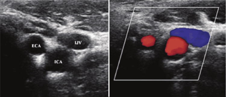
Principle of ultrasound : When a sound wave strikes an object it is reflected, by measuring this echo it is possible to determine the distance of the object as well as the size, shape and consistency of the object. bodies, including solids and liquids. General Principle of the Doppler Effect: Doppler ultrasound is a special ultrasound technique that measures the direction and speed of blood cells as they move through the vessels. The movement of blood cells causes a change in the reflected sound waves (known as the Doppler effect). A computer will collect and process it to create charts or color images that represent the blood flow through the blood vessels.

Hình ảnh siêu âm Doppler động mạch cảnh
4. Procedure of carotid Doppler ultrasound
Prepare the patient : The patient is dressed in such a way that the ultrasound procedure is easy. Remove neck jewelry. Explain to the patient the technique and timing of the procedure (the procedure takes place over a period of 30 minutes). Equipment: Color ultrasound machine with flat transducer, patient bed. The person performing the procedure is a doctor specializing in diagnostic imaging. Procedure: The patient lies supine on the ultrasound bed, level with the doctor's sitting position, with a pillow on the neck area so that the head is tilted down, so that the carotid artery is exposed as wide and as long as possible, during the procedure. The patient may be asked to tilt to the right or to the left so that the doctor's sonographic manipulation is easy. There is a gel that is sprayed into the survey area so that the ultrasound probe is in contact with the gel to capture a clear carotid image. This ultrasound process lasts between 30 - 45 minutes.
To register for examination and treatment at Vinmec International General Hospital, you can contact Vinmec Health System nationwide, or register online HERE.


