This is an automatically translated article.
The article was written by Doctor of Radiology, Department of Diagnostic Imaging - Vinmec Central Park International General Hospital.Complications of myocardial infarction are very serious and can be life-threatening. Doppler echocardiography is a measure to help evaluate complications of myocardial infarction patients to help doctors make appropriate treatment options and prognosis.
1. Complications of myocardial infarction
1.1 What is a Heart Attack? Myocardial infarction is the death of the heart muscle due to ischemia. Most cases of necrosis are related to occlusion or near complete occlusion of the coronary lumen.
When having an acute myocardial infarction, the patient has some characteristic signs such as pain behind the sternum or in front of the heart, pain on the left shoulder and the inside of the left hand, sudden onset of heart attack, lasting more than 20 minute. Pain may radiate to the patient's neck, chin, shoulder, back, right hand or epigastrium. In addition, patients may also experience some other symptoms such as sweating, shortness of breath, vomiting or nausea, cold extremities, confusion, pale skin, etc. due to cardiovascular collapse or low blood pressure. .
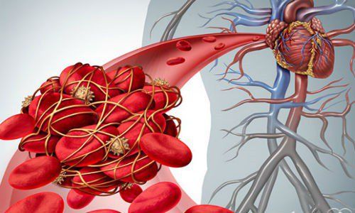
1.2 Common Complications of a Heart Attack Arrhythmia: An irregular heartbeat, which may be faster or slower than average. Common arrhythmias include supraventricular arrhythmias (such as sinus bradycardia, sinus tachycardia, paroxysmal supraventricular tachycardia, atrial fibrillation) and ventricular arrhythmias (ventricular fibrillation, ventricular tachycardia),...; Pump failure: Left ventricular failure, systolic failure and diastolic failure. The most severe complication of pump failure is cardiogenic shock, showing cardiovascular collapse, oliguria or anuria, pale extremities, impaired consciousness,...; Atrial block most: Common in people with posterior - inferior myocardial infarction, can occur suddenly, high mortality rate; Mechanical complications: Rupture of the left ventricular free wall; rupture of the interventricular septum forming a ventricular septal defect; rupture of the papillary muscle in the valve leaflet causes valve prolapse, acute mitral regurgitation; Thromboembolic complications: Increases the risk of recurrent myocardial infarction, causing extensive necrosis or new myocardial necrosis, systemic embolism, pulmonary embolism; Other early complications: Acute pericarditis, sudden death; Late complications: Dressler syndrome; fever, chest pain when taking a deep breath; leukocytosis, increased erythrocyte sedimentation rate; ventricular aneurysm; angina; heart failure ; sudden death; periarthritis of the shoulder, ... after myocardial infarction.

Đau thắt ngực là biến chứng hay gặp của nhồi máu cơ tim
2. The role of Doppler echocardiography in assessing complications after myocardial infarction
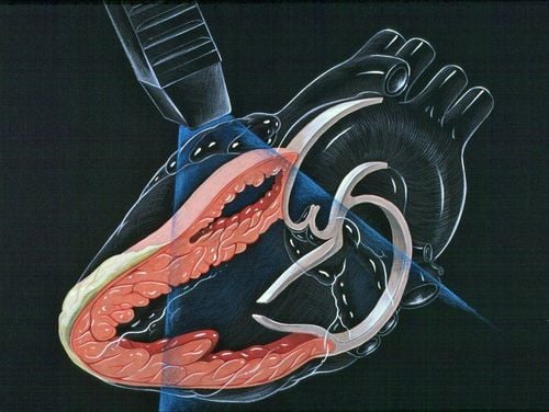
2.1 Advantages of Doppler echocardiography In patients with myocardial infarction, Doppler ultrasound helps diagnose, determine the location and extent of myocardial infarction, detect complications after myocardial infarction and provide an appropriate prognosis. This method possesses many advantages such as non-invasive, quick at the bedside, reproducible without danger, high accuracy, low cost. At the same time, it provides anatomical information, hemodynamics, and the status of blood flow in the circulatory system, which is very useful for the physician's diagnostic evaluation.
2.2 Types of Doppler Echocardiography Cardiac Doppler Echocardiography includes:
Two-dimensional (2D) Echocardiogram; Echocardiography TM; Doppler ultrasound of myocardial tissue; Ultrasound - Pulsed Doppler; Continuous Doppler and Color Doppler.
2.3 How does Doppler echocardiography assess complications after myocardial infarction? There are 3 groups of complications after myocardial infarction that can be detected through Doppler echocardiography: mechanical complications, ventricular thrombosis and pericardial effusion. Specifically:
Pericardial effusion: Application of 2D ultrasound through the chest wall to assess the amount of fluid, the location of the fluid and monitor the change over time during the treatment;
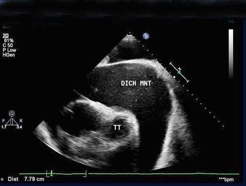
Hình ảnh tràn dịch màng ngoài tim trên siêu âm
Heart aneurysm: On ultrasound, the image of an aneurysm includes the aneurysm neck (the junction between the normal ventricular wall and the aneurysm of the heart) with thickened walls and normal movement; Aneurysms are characterized by thin walls, increased brightness. On ultrasound, the aneurysm area is flared, movement is reversed during systole. Often, the sound coil and thrombus in the aneurysm are common; Pseudoaneurysm: On 2D ultrasound, a narrow perforation, communicating with the heart chambers, has a relatively sharp border. During systole, the pseudocyst dilates. Pulsed Doppler ultrasound when the window is placed on the neck of the aneurysm will yield a double-peaked positive wave. On color sonography, the flow through the neck of the aneurysm is seen, the flow is usually bidirectional, there is eddy blood flow in the pseudoaneurysm and thrombus can be seen in the aneurysm; Thrombosis: 2D ultrasound can detect thrombus (abnormally dense acoustic structures in the heart chambers, often at the site of myocardial infarction, appearing on at least 2 planes, continuously throughout the entire cycle. cardioversion); Interventricular septal perforation: In many cases, 2D ultrasound can show the hole, usually in the septum adjacent to the apex of the heart. However, there are some cases where the hole cannot be clearly seen on 2D ultrasound. In this case, color ultrasound is performed to detect the stoma. Doppler ultrasound can show high-speed spectrum during systole with left-right direction, estimate pulmonary artery pressure, shunt ratio; Mitral regurgitation: The signs of this post-myocardial infarction complication can be seen indirectly on IV and 2D ultrasound such as left ventricular dilatation, left atrium. Doppler ultrasound and color ultrasound can be used to assess the extent of mitral regurgitation. Myocardial infarction causes many different complications, and Doppler ultrasound is the leading diagnostic method because of its simplicity, high accuracy, and safety for the patient. Therefore, when there are any warning signs of this condition, the patient should go to the doctor immediately so that the doctor can appoint an appropriate diagnosis, thereby having the best treatment plan.
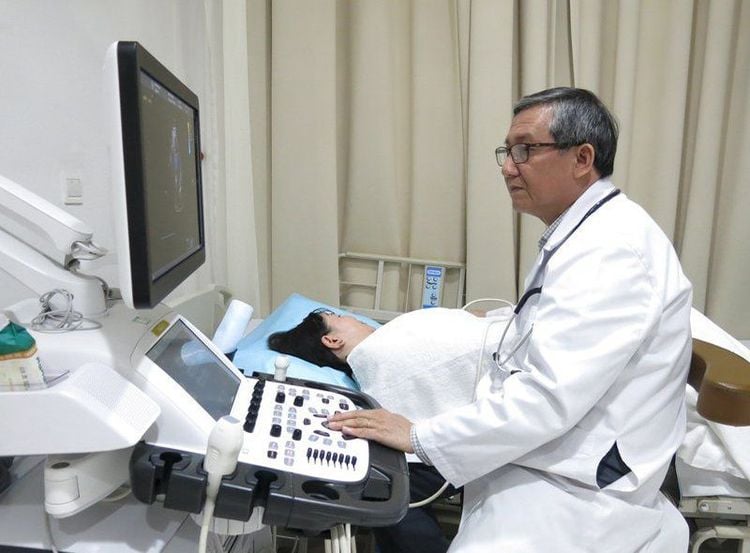
Kỹ thuật siêu âm tim cần được thực hiện bởi bác sĩ chuyên khoa giàu kinh nghiệm
To protect heart health in general and detect early signs of cardiovascular disease, customers can sign up for Cardiovascular Screening Package - Basic Cardiovascular Examination of Vinmec International General Hospital. The examination package helps to detect cardiovascular problems at the earliest through tests and modern imaging methods. The package is for all ages, genders and is especially essential for people with risk factors for cardiovascular disease.
Please dial HOTLINE for more information or register for an appointment HERE. Download MyVinmec app to make appointments faster and to manage your bookings easily.


