This is an automatically translated article.
The article was professionally consulted by Specialist Doctor II Nguyen Quoc Viet - Department of Medical Examination & Internal Medicine - Vinmec Danang International General Hospital. The doctor has more than 20 years of experience in the examination and treatment of cardiovascular diseases and Cardiovascular Interventions (Including angiography, dilation, stenting of coronary arteries, renal arteries...), placing temporary pacemakers , forever...Cardiac enlargement, dilated cardiomyopathy, or in medicine called dilated cardiomyopathy, is a disease of the heart muscle, usually starting from the heart's main pumping chamber, the left ventricle, losing its firm elastic structure. The disease may initially cause no symptoms, but once symptoms are present, they progress rapidly, have a severe prognosis, and eventually lead to potentially life-threatening events.
1. What is an enlarged, dilated heart (dilated cardiomyopathy)?
Cardiac enlargement, dilated cardiomyopathy, the medical term is Dilated cardiomyopathy, which is characterized by left ventricular dilatation with systolic dysfunction and is not caused by ischemic heart disease, hypertension, or congenital heart disease. birth or heart valve disease. However, in clinical practice the concept of ischemic cardiomyopathy, hypertensive cardiomyopathy is still used.The disease can affect people of all ages, including infants and children, but is most common in men between the ages of 20 and 50.
Common causes of cardiomegaly are:
Toxins: alcohol, cocaine, cancer chemotherapy... Genetic or familial Infections: HIV, CMV, diphtheria Metabolic abnormalities Chemical: hypothyroidism, hyperthyroidism, renal failure, Cushing, Vitamin B1 deficiency, hypocalcemia, hypophosphatemia, hemochromatosis Inflammatory: systemic lupus erythematosus, scleroderma Idiopathic dilated cardiomyopathy
2. What are the symptoms of dilated cardiomyopathy?
Patients with enlarged heart, dilated heart chambers often have symptoms of heart failure but often progress slowly, maybe months or years without symptoms. When present, the onset is usually progressive heart failure, in addition to thromboembolism or syncope.Different from common cardiovascular diseases, dilated cardiomyopathy can occur at any age, but it is more common in middle-aged people, more men than women. A small number of patients have a family history with a diagnosis of dilated cardiomyopathy or alcohol or cocaine addiction or a recent viral infection.
Patients will go to the doctor when they find that their body is tired, getting weaker and weaker, shortness of breath with exertion, shortness of breath when lying down, paroxysmal nocturnal dyspnea. Some cases have symptoms of right heart failure such as weight gain, abdominal bloating, nausea, right upper quadrant pain due to hepatomegaly, ascites...
When examining, the doctor will detect typical symptoms of heart failure. Chronic low-output heart failure with normal or low blood pressure, narrow BP gradient, and staggered pulse in severe heart failure. When the patient is in the lying position, the signs are easily detected such as enlarged liver, distended neck veins, edema of the limbs, rales in the lungs. When palpating the heart, the heart area is enlarged, the apex is deviated and the heart has a split T2 sound, T4 is a precursor to severe heart failure. In addition, a systolic murmur due to functional regurgitation is heard in the mitral and tricuspid valves.

Bệnh có thể xảy ra ở mọi lứa tuổi nhưng thường gặp người trung niên
3. What diagnostic tests for dilated cardiomyopathy should be done?
Similar to other cardiovascular diseases, before the diagnosis of dilated heart disease, the patient will be assigned to perform the following tests:Chest X-ray: Enlarged heart, pulmonary congestion, pleural effusion Pulmonary
Electrocardiogram : Although there are no typical signs of dilated cardiomyopathy, ECG results may also show sinus tachycardia, atrial fibrillation, and ventricular extrasystoles. Left bundle branch block, left axis of displacement, left ventricular enlargement and may have myocardial infarction-like appearance in dilated cardiomyopathy. For Holter ECG can record ventricular and supraventricular arrhythmias...
Echocardiography: This is an effective method to diagnose and monitor dilated cardiomyopathy when the first thing to do is possible. Exclude valvular heart disease, pericardial disease, congenital heart disease. Next, echocardiography will easily detect left ventricular dilatation, right ventricular dilatation, and impaired left ventricular systolic function with low ejection fraction associated with mitral regurgitation. Pericardial effusion can be detected. Finally, there is a sign of regional dyskinesia when it is usually uniformly reduced movement of the walls. If there is a localized movement disorder, the prognosis is much better.
Dobutamine stress echocardiography: Unlike conventional ultrasound, inotropic drugs with dobutamine will help in the differential diagnosis of ischemic cardiomyopathy and idiopathic dilated cardiomyopathy.
Cardiovascular magnetic resonance imaging: This is a new means of accurately assessing left ventricular function, helping to replace coronary angiography in diagnosing and excluding ischemic causes based on the pattern of late increased signal distribution. with gadolinium.
Coronary angiography: This is an interventional method to help diagnose and exclude coronary artery disease or suspected coronary artery disease when non-invasive test results are inconclusive
Other tests help Excluding secondary causes such as thyroid hormone, kidney function, blood calcium level, blood phosphate, serum Fe, HIV... BNP or pro-BNP will help diagnose heart failure and patient prognosis.
4. How to treat dilated heart disease?

Bỏ rượu, thuốc lá là biện pháp điều trị không thuốc của bệnh tim to giãn buồng tim
Non-drug measures: lose weight, limit salt in the diet, quit smoking, alcohol, stimulants... Medication measures: with the leading drug classes are ACE inhibitors to reduce the load on the heart, prolong survival in patients with heart failure, can be replaced by receptor blockers when ACE inhibitors are not tolerated. Besides, beta blockers, aldosterone antagonists will reduce the mortality rate of patients with heart failure and use diuretics to improve symptoms in cases of volume overload. If patients with severe heart failure develop, add vasopressors such as dopamine, dobutamine. Finally, consider the indication for heart transplantation in cases of end-stage heart failure and unresponsive to medical treatment.
5. What are the complications and prognosis of dilated cardiomyopathy?
Rối loạn nhịp tim là một trong những biến chứng của bệnh tim to giãn buồng tim
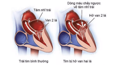
Heart failure: Poor blood flow from the left ventricle can lead to heart failure. At this point, the heart can no longer supply the blood the body needs to function properly. Heart valve regurgitation: Dilated heart chambers can make it harder for your heart valves to close properly, causing backflow every time your heart needs to contract to expel blood. Generalized edema: Excess water is not eliminated by the kidneys due to decreased cardiac output. Fluid buildup causes swelling in the legs and fluid retention in the lungs and abdomen. Cardiac arrhythmias: Changes in heart structure and changes in pressure on the heart's chambers can contribute to disturbances in electrical conduction pathways. The risk of cardiac arrest and sudden death increases many times over. Blood clot formation: Blood pooling in the left ventricle due to enlarged heart chambers can lead to the formation of blood clots. If they are accidentally expelled, they can block blood vessels and cause ischemia to vital organs, causing stroke, heart attack, or other organ damage. Prognosis of dilated cardiomyopathy usually has a 5-year mortality rate of 20%. Factors contributing to a severe prognosis are age over 55 years, auscultation with T3 sound, ECG with endocardial conduction block, degree of chamber dilation, degree of heart failure, degree of reduction in ejection fraction and cardiac output. echocardiography and life-saving ventricular arrhythmia events.
In summary, dilated cardiomyopathy or dilated cardiomyopathy is the final complication of some disease or is the primary disease in the first place. Regardless of the cause, the disease often has severe symptoms, the patient's independent living function is severely impaired, and the long-term prognosis is poor. Therefore, it is necessary to detect dilated cardiomyopathy early and actively treat it at the very beginning in the hope of maintaining long-term quality of life for patients.
To protect cardiovascular health in general and detect early signs of myocardial infarction and stroke, customers can sign up for Cardiovascular Screening Package - Basic Cardiovascular Examination of Vinmec International General Hospital . The examination package helps to detect cardiovascular problems at the earliest through tests and modern imaging methods. The package is for all ages, genders and is especially essential for people with risk factors for cardiovascular disease.
Please dial HOTLINE for more information or register for an appointment HERE. Download MyVinmec app to make appointments faster and to manage your bookings easily.