This is an automatically translated article.
This article was professionally consulted by a Doctor of Radiology, Department of Radiology - Vinmec Central Park International General Hospital.For intervention and treatment of peripheral vascular diseases, angiography and direct embolization are often used through the skin.
1. What is digitizing background removal and direct percutaneous embolism?
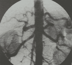
Digitizing background erasure and direct percutaneous embolization is an interventional method to treat peripheral vascular malformations. Digital background erasure and direct percutaneous embolization can be applied alone or in combination with traditional embolization techniques.
This method will be performed by inserting a needle into the malformation, then angiography with iodine contrast to assess the hemodynamic status of the lesion. Finally, inject drugs or embolization materials to complete the steps of the procedure.
2. Indications, contraindications for digital background removal and direct percutaneous embolism
2.1 Designation
Usually this method will be indicated for cases of arteriovenous malformation, venous malformation or hemangioma.
2.2 Contraindications
Allergy to iodinated contrast agents. Inflammation, infection, and necrosis of the skin and soft tissues of the area intended for direct puncture. Pregnant. Severe and uncontrolled coagulopathy (prothrombin <60%, INR >1.5, platelet count <50 G/l).

Phụ nữ có thai chống chỉ định thực hiện thủ thuật này
3. Preparing for the procedure of digital imaging to erase the background and cause direct embolism through the skin
3.1 Participants and tips
Specialist. Auxiliary physician. Optical technician. Nursing. Doctor, anesthesiologist (if the patient cannot cooperate).
3.2 Means used in digitized imaging to erase the background and cause direct percutaneous embolism
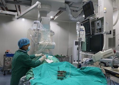
Dedicated electric pump. Digital background removal angiography (DSA). Lead vest, apron, X-ray shielding Film, film printer, image storage system.

Máy chụp mạch số hóa xóa nền (DSA) không thể thiếu trong quá trình chụp số hóa xóa nền và gây tắc mạch trực tiếp qua da
3.3 What drugs should be prepared for the process of digital imaging to erase the background and cause direct embolism through the skin?
Digital background removal and direct percutaneous embolization requires the use of the following drugs:
Local anesthetic. Pre-anesthesia and general anesthetic (if there is an indication for anesthesia). Water-soluble iodine contrast agent. Antiseptic solution for skin and mucous membranes. Anticoagulants. Anticoagulant neutralizer.
3.4 Common medical supplies to prepare
Syringe for electric pumps. Syringe 1; 3; 5, 10; and 20ml. Distilled water or physiological saline. Gloves, shirt, hat, surgical mask. Cotton, gauze, surgical tape. Medicine box and first aid kit for contrast drug accidents. Aseptic intervention kit: knife, scissors, tongs, 4 metal bowls, bean tray, tool tray.

Cần chuẩn bị các vật tư y tế thông thường trước khi thực hiện thủ thuật
3.5 Special medical supplies for digitized background removal and percutaneous embolization
Pulse needle, butterfly needle. Y-connector set. 3-prong lock. Instrumentation kit.
3.6 Embolic materials
Bio-foam (gelfoam,...). Bio-glue (Histoacryl, Onyx,...). Absolute alcohol.
4. Steps to conduct digital imaging to erase the background and cause embolism directly through the skin
Step 1: Open the way into the vessel
Local anesthetic. Use an appropriately sized needle to poke the lesion. Needle puncture can be performed under ultrasound and/or DSA guidance. Step 2: Angiography to assess the damage
Connect the puncture needle to the connecting wire. Perform systemic angiography to assess the hemodynamic status of the lesion and adjacent vascular sites. Step 3: Intervention therapy
Each morphological and hemodynamic characteristics of the lesion will have decisions to choose different embolization materials, some materials can be used such as metal spirals (Coils), bio glue (nBCA, Onyx), fiber (Tromboject) or Ethanol. Insert embolization material into the lesion for embolization. If the embolization material is a liquid solution (bio-glue) and the lesion has a large flow rate and has drainage veins, it is necessary to incorporate a tourniquet of the vein above the lesion (proximal limb). Step 4: Evaluation after intervention
Angiography assesses circulation after recanalization. Withdraw the puncture needle, gently apply pressure to the puncture area

Các bước tiến hành chụp số hóa xóa nền và gây tắc mạch trực tiếp qua da
5. Evaluating the results of digitizing scans that erase the background and cause direct embolism through the skin
Digital background erasure and direct percutaneous embolization were considered successful when all malformations were removed from the circulation and no flow signal remained.
At the same time, the branches of the arteries supplying blood to the downstream area and the draining veins are still circulating normally.
6. Things patients need to pay attention to when performing the procedure
First, you must have a good understanding of the procedures in digitizing background removal and direct percutaneous embolization so that you can coordinate with your doctor. Within 6 hours before the procedure, the patient needs to fast, and can only drink an amount of water not exceeding 50ml.
In the intervention room, the patient lies on his back, with a machine to monitor breathing, blood pressure, pulse, electrocardiogram, SpO2. Disinfect the skin, then cover with a sterile, perforated cloth. If the patient is too excited, cannot lie still, it is necessary to give sedation or take appropriate measures.
Any questions that need to be answered by a specialist doctor as well as if you have a need for examination and treatment at Vinmec International General Hospital, please book an appointment on the website to be served.
Please dial HOTLINE for more information or register for an appointment HERE. Download MyVinmec app to make appointments faster and to manage your bookings easily.


