This is an automatically translated article.
Posted by Resident Doctor, Master Tran Duc Tuan - Department of Diagnostic Imaging - Vinmec Central Park International General Hospital
Digital erasure scan, also known as DSA scan of the aorta and thoracic aorta, allows to show the structure and detect vascular lesions in the chest area so that the doctor will make a treatment plan. disease suitable for each patient's condition
1. What is thoracic aortic background digitization?
Digital erasure scan, also known as DSA scan of the aortic arch and thoracic aorta, is an imaging technique using iodinated contrast, thereby displaying the anatomical structure of the aortic arch and arteries. Larger sources arise from the aortic arch such as the thoracic aorta, the left subclavian artery, the left carotid artery, and the brachiocephalic artery.
2. Digital angiography erasing the thoracic aorta is indicated for whom?
Digitization of thoracic aortic arch and thoracic aorta is indicated for the following cases:
Congenital disease and diseases related to the aortic arch, thoracic aorta and its branching arteries such as: thoracic aortic aneurysm, thoracic artery stenosis, arteriovenous catheterization, vascular tumor, underdeveloped vessel, ... Suspect trauma to the chest area causing vessel damage. Suspected aortic abnormality causing cerebral ischemia. Suspect a tumor in the chest that compresses or invades the blood vessels. Assess the arterial blood supply to the tumor. Serving electro-optical intervention.

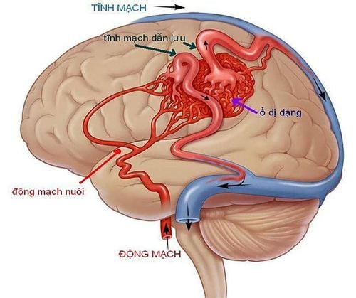
Bệnh nhân thiếu máu não do động mạch chủ gây ra được chỉ định thực hiện
3. How is the thoracic aortic background digitized scan performed?
DSA scan of the aorta and thoracic aorta is performed with the following steps:
Step 1: Anesthetize the patient by asking the patient to lie supine on the imaging table, setting up an intravenous line, local anesthetic. General anesthesia can be given before the procedure by injecting pre-anesthesia in cases where the patient is agitated, fearful or unable to cooperate (children under 5 years old).
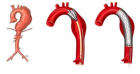
Chụp số hóa xóa nền cung động mạch chủ và động mạch chủ ngực giúp phát hiện những tổn thương mạch như phình động mạch chủ ngực
Step 2: Use the Seldinger technique to insert the catheter from the femoral artery. Disinfect and anesthetize the needle puncture site to insert the catheter. If access from the femoral artery is not possible, alternative access from the axillary, brachial, or radial artery may be selected. Step 3: Conduct aortic arch and thoracic aorta. For the aortic arch, insert the catheter guide into the lumen up the abdominal aorta and the thoracic aorta, then insert the catheter tip through the aortic arch to reach the aortic valve sinus. Note, keep 2cm from the valve. For the thoracic aorta, the catheter tip should be retracted. Step 4: Use a pump to inject about 30-40 ml of iodinated contrast agent with high pressure of 800 PSI, pumping speed is about 15-20 ml/s. Note, it is necessary to avoid the movement of the heart rate to interfere with the image, then, take continuous DSA. For the arch of the aorta, imaging should be done anterolaterally to the left and focus on the arch of the aorta, as well as the brachiocephalic artery, subclavian artery, and left carotid artery, which are arteries arising from the arch. aorta. For the thoracic aorta, the image can be straight or angled forward, to the left, and focuses on the thoracic aorta. Step 5: Finish the capture process when the requirements are met, proceed to withdraw the tube. After that, stop bleeding for the patient by applying direct pressure on the needle puncture site for 15 minutes, using a special kit to close the lumen or applying pressure for 6 hours. The right imaging results clearly show the structure of the ascending and descending aorta, the aortic arch and the great branches of the aorta originating from the aortic arch such as the brachiocephalic artery, the left common carotid artery, the left common carotid artery, the cephalic artery, the left common carotid artery, the cephalic artery, the left common carotid artery, the cephalic artery, the left common carotid artery, the cephalic artery, the left common carotid artery, the cephalic artery, the left common carotid artery, the cephalic artery, the cephalic artery, the left common carotid artery, the cephalic artery, the cephalic artery, the left common carotid artery, the cephalic artery, the left common carotid artery, the cephalic artery, the left common carotid artery, the cephalic carotid artery, the cephalic artery, the left common carotid artery, the cephalic artery, the left common carotid artery, the cephalic artery, the left common carotid artery, and the cephalic artery. left subclavian pulse. From there, it is possible to detect suspected lesions, if any.
Digital angiography erases the background of the aortic arch and thoracic aorta is a modern imaging technique used to diagnose and detect vascular lesions, thereby having appropriate and timely treatment. .
Abdominal aortic angiography is a modern imaging technique that helps to detect lesions in the abdominal aorta, thereby providing appropriate treatment.
Digital angiography erases the background of the aortic arch and thoracic aorta is a modern imaging technique that helps doctors detect the location and extent of lesions of the disease. However, for accurate results, patients should choose reputable addresses with modern DSA scanners, and at the same time, it must be performed by qualified technicians and doctors.

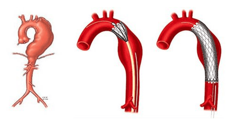
Chụp số hóa xóa nền động mạch chủ ngực giúp phát hiện những tổn thương ở vùng động mạch chủ bụng
Vinmec International General Hospital has applied digital imaging technology to erase the background in examination and diagnosis of many diseases. The process of digitizing and erasing the background at Vinmec is carried out methodically and according to the standards of the process by a team of highly skilled doctors and modern machinery, thus giving accurate results, making a significant contribution. in determining the disease and the stage of the disease for timely treatment.
Especially now, to improve the quality of medical examination and treatment services, Vinmec continues to deploy many perfect medical services to bring convenience and satisfaction to customers.
If you have a need for medical examination by modern and highly effective methods at Vinmec, please register here.